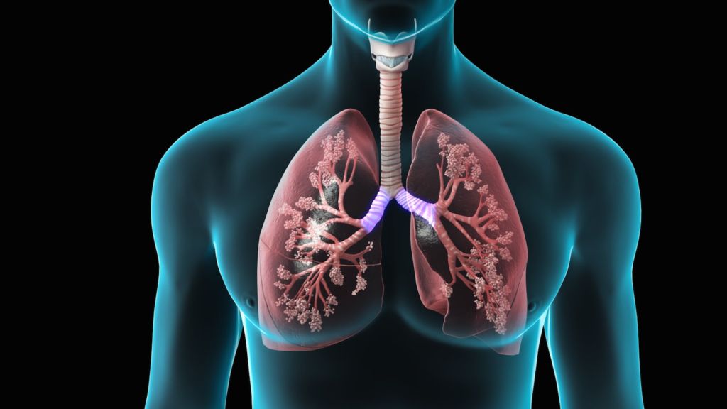
Coating the laminar surface of all endothelial cells is endothelial surface layer. This layer was discovered two decades ago, but its imaging is still found to be difficult. Successful endothelial surface layer imaging would be medically beneficial, as it could quantify inflammatory conditions. In this particular case, researchers examine pulmonary tissues of the lung. Image courtesy of YouTube.
A joint team of researchers from the Korea Advanced Institute of Science and Technology and the Seoul National University have achieved in vivo imaging of the endothelial surface layer. The endothelial surface layer is located on the extracellular surface of endothelial cells which coat blood vessels. Its function is to serve as a form of protection. This layer consists of glycocalyx, a cellular coating of glycoproteins and glycolipids, and bound plasma constituents. Until now, endothelial surface layer imaging has been a difficult task. To tackle this challenge, the researchers custom designed a laser scanning confocal microscope and a pulmonary imaging window chamber. At the conclusion of the experiment, they were able to visualize endothelial surface layer damage in a model of pulmonary sepsis.
Why is endothelial surface layer imaging important in the medical field?
Recent research suggests that the endothelial surface layer is damaged in inflammatory conditions. Such inflammatory conditions include sepsis, a dangerous complication of an infection. Currently, early sepsis is difficult to diagnose, and is only noticed in its late stages, when organ dysfunction occurs. During an inflammatory cascade, endothelial cells may shed their glycocalyx coating. Therefore, endothelial surface layer imaging can help quantify damage done to endothelial cells or to detect harmful inflammatory conditions.
Ex vivo imaging has so far proved unsuccessful, as the thin gel-like layer is damaged when fixed. Additionally, in vivo imaging is made difficult as the thickness of the layer is determined by the type of blood cell passing through. Leukocytes, which play a major role in inflammation, damage glycocalyx while erythrocytes, red blood cells, do not. Because of this, the team of researchers focused their experiments on pulmonary endothelial tissue in the lungs, as it has a large erythrocyte flow.
How were experiments run?
For the experiments, researchers built a laser scanning confocal microscope. The wavelengths output by the microscope to be used as excitation light sources were 488 nm, 561 nm, and 640 nm. To obtain 30 Hz video-rate laser scanning, a rotating 36-faceted polygonal mirror was implemented. Finally, multi-color fluorescence imaging was achieved via the use of three photomultiplier tubes with bandpass filters.
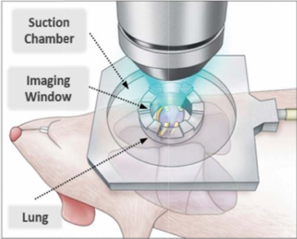
During experimentation, researchers performed a thoracotomy, an incision on the chest wall, on mice under anesthesia. This exposed the pleura, the membranes which covers the lungs. Following this simple surgery, the mouse was hooked up to the imaging system, as shown in the image below. Fluorescein isothiocyanate (FITC) conjugated dextran dye was inserted into the mouse via catheter to allow for fluorescent visualization.

The image above details how the imaging device is placed in relation to the mouse subject. Suction pressure is applied to minimize noise caused by motion during imagine. Image courtesy of Biomedical Optics Express.
What were the findings of this experiment?
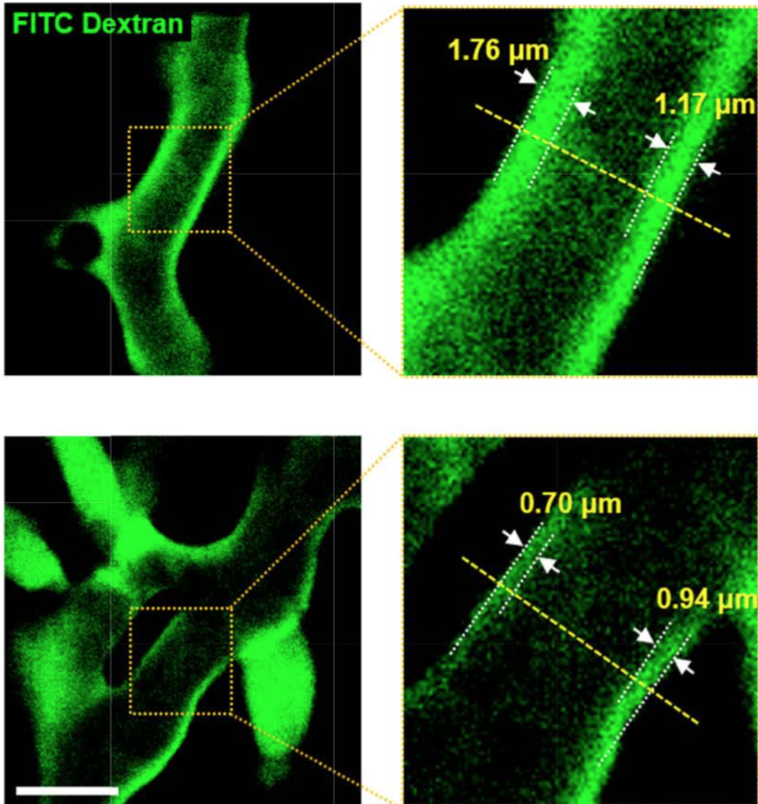
The researchers were successful in imaging the endothelial surface layer of pulmonary tissue in live mice. These images can be seen below. By averaging together the many frames of the obtained videos, researchers could easily identify the lumen of the capillary, where the erythrocytes flow through, as well as the endothelial surface layer through the use of the green FITC dye. Images obtained from the pulmonary tissue of a mouse with a pulmonary inflammation support research that the endothelial surface layer is damaged by inflammation.

The graphics above show obtained images from the experiment. On the top row, the capillary of a healthy mouse can be seen. The green fluorescent dye indicates endothelial surface layer. The bottom layer shows the capillaries of a mouse with a pulmonary inflammation. As can be seen, the endothelial surface layer has degraded. Image courtesy of Biomedical Optics Express.
According to the researchers, this is the first time such images of intravital pulmonary endothelial surface layer have been made by analyzing the flow of erythrocytes.
Where does the research go from here?
Obtaining such clear images of endothelial surface layer and visualizing damage to it is a large first step. By expanding trials to include other mammals and other types of tissues, more progress can be made. A foreseeable challenge to overcome is imaging tissues with a large, natural leukocyte concentration. As stated earlier, leukocytes can damage and cause non-homogenous endothelial surface layer, which is more difficult to image. Another possible improvement to be made would be making the technology less invasive. In terms of medical use, this technology can go on to diagnose sepsis based on endothelial surface layer damage. This is vital in the medical field as often times such complications can go unnoticed. By making the imaging system less invasive, doctors wouldn’t require large surgeries to diagnose possibly dangerous inflammatory conditions.
To read more about this ground-breaking experiment, the paper can be found here.
