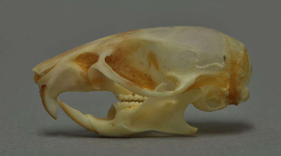
Calvarial fractures refer to defects in the flat bones that make up the skull. Raman spectroscopy has been identified as a technique that can be used to image the healing process of these fractures. This is a novel calvarial fracture imaging technique. Image Courtesy of Bioengineering Today.
Scientists from the City University of Hong Kong’s Department of Physics revolutionized calvarial fracture imaging through the use of Raman spectroscopy. By allowing these defects to be imaged, the healing process can be studied. Specifically, bone composition throughout the healing process can be quantified and measured. This new imaging technique is a safer procedure that will not disturb the actual bone as it is self-repairing itself.
What are calvarial fractures?
The term calvaria refers to the skullcap. It is made up of bones that protect the cranial cavity, which houses the brain, at the top of the skull. Calvarial fractures refer to defects in these bones. Like many bone fractures, these fractures heal by themselves in most cases, and often without complications. The healing process involves the formation of fragile soft callus, which is later replaced by hard callus, a woven bone material.
In the case of calvarial fractures, there are two distinct types: critical and subcritical. Critical defects do not heal naturally, and frequently require surgery. Subcritical defects do heal spontaneously in the majority of cases. This new calvarial fracture imaging technique is focused on rats with subcritical calvarial defects.
How have these fractures previously been imaged?
In clinical settings, a skull fracture is first diagnosed via a CT scan. This involves the use of X-rays to take multiple images of the skull and brain. A CT scan is very efficient at identifying the placement, size, and severity of a skull fracture.
After the fracture is diagnosed, the healing process can be tracked through more CT scans and other radiographic imaging as well as clinical examinations. Unfortunately, these methods cannot give any detailed information on the changes in the mineral and protein composition of the healing bone. Information on the composition of the bone is useful to researchers as it can allow them to asses the strength of the new bone as well as the rate of healing.
How does Raman spectroscopy help image bone fractures?

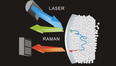
The above graphic shows a simplified overview of Raman technology. A charge coupled device measures and collects the light scattered after a laser is fixated on the sample. Image Courtesy of OrthoStreams.
Raman spectroscopy is a widely-used imaging technique similar to infrared absorption spectroscopy. It requires shining a light source on the subject and measuring the scattering of light. Subsequently, chemical property determination and molecular identification can be gained from the data of Raman spectroscopy. Developed in 1928, Raman spectroscopy is a clever analytical tool that relies on the unique properties of molecular bonds that vibrate at peculiar wavelengths. As a result, each molecule contains a specific spectroscopic marker that can be observed.
For samples of bone, Raman spectroscopy can characterize the mineral lattices as well as the protein matrices. This provides important data to researchers who are concerned about the mineral and protein makeup of healing bone. Raman spectroscopy has previously been used in bones’ study to measure effects of mechanical load, especially with so-called femur and tibia fractures. However, it has not yet been used to track the healing of calvarial fractures. Studying the bone composition using Raman spectroscopy will allow researchers to do just that.
Now, you may ask, how did researchers test calvarial fracture imaging through Raman spectrometry?
The test subjects for the experiments run by the researchers were rats. While anesthetized, the skull was exposed and a 1 mm wide and 200 μm deep defect was made with a metal burr drill. The defect healed for 7 or 14 days before researchers euthanized the rats. After researchers harvested the bone, a control defect was also made next to the original defect.

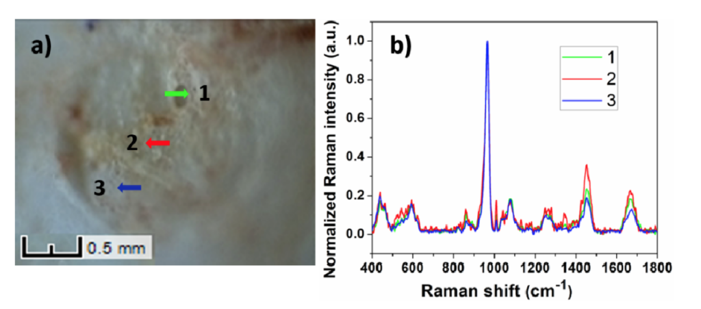
The image on the left shows a defect on a sample of bone harvested after 14 days of healing. The green arrow points to an area of hard callus. The red arrow points to an area of soft callus. Finally, the blue arrow points to an area of healed bone. The graph on the rights shows the corresponding spectra. Image Courtesy of Biomedical Optics Express.
The skull samples were then imaged using Raman spectroscopy. The laser used was a 785 nm, 250 mW excitation laser. The laser was focused on 10 distinct sites on each sample to get 10 Raman spectra. This increased data obtained and improved coverage of each fracture. A high sensitivity open electrode charge coupled device collected Raman scattering. This charge coupled device had 1024 x 256 pixels. The spectra were processed with the Vancouver Raman Algorithm before being smoothed and filtered. Researchers were mostly concerned with mineral to matrix ratio, carbonate to phosphate ratio, and crystallinity.
How successful was this new method of calvarial fracture imaging?
After analysis of the data, researchers could identify soft callus, a cartilaginous material, hard callus, the calcified cartilaginous material and woven bone. These three distinct stages of healing were distinguished through changes in the peaks in the Raman spectra, as shown in the image in the previous section. It was noted that soft callus formation peaked around day 7 following injury. Hard callus development peaked around day 14. As hard callus formation progressed, woven bone replaced the callus. The area of the defect becomes more rigid as healing occurred.
To summarize, this technique was highly successful. It provided an easy way to track the progression of healing of calvarial fractures that incurs minimal, if any, damage to the bone while providing detailed results. Advances like this might just create a breakthrough in the medical field allowing doctors to develop progressively less invasive healing procedures.
To read more about this calvarial fracture imaging technique, check out their original paper which can be found here.
