The cornea protects the eye from debris and germs from the environment and accounts for roughly two-thirds of the eye’s focusing power. Corneal imaging is critical in diagnosing and determining treatment for diseases and eye-related health problems, as well as monitoring their progression. The cornea can be difficult to image since it appears clear to most visible and infrared light. Despite the difficulty, there are many methods for imaging the cornea. Methods explored below include selected forms of topography, tomography, and microscopy.
Corneal Topography
Corneal topography profiles the cornea’s surface like cartographers use topography to profile landmasses. It uses reflection to profile the anterior surface of the cornea. Topography is used for preliminary imaging of the cornea to determine the fit for surgeries and for postoperative assessment.
Placido Discs
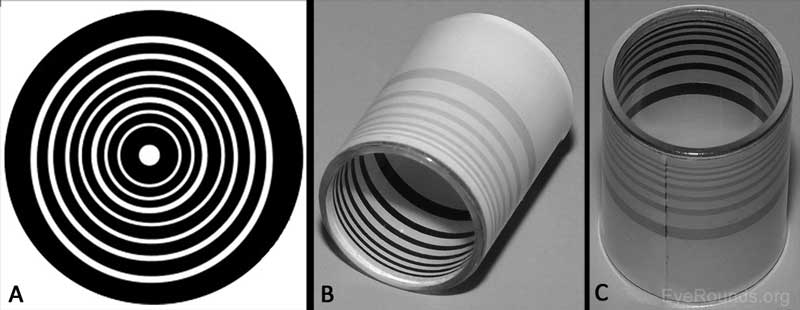
Placido discs were some of the first tools to be used in topography. A Placido disc is an image of alternating black and white concentric circles. An image of the disc would be projected onto the cornea and the resulting pattern of the light reflected could then be assessed. Distortion of the resulting image could indicate irregularities of the cornea. For instance, a resulting image with irregular spacing between the disc rings could indicate a steeper curve of the cornea surface in that area. A manual assessment tool you may have seen at the ophthalmologist using Placido discs is known as a keratoscope (often used to check for astigmatism or dystrophies). Some more advanced topography methods still implement the Placido disc method but supplemented with computer analysis.

Pictured in (A) is an example of what a Placido disc looks like. (B) and (C) are old versions of keratoscopes. Courtesy of University of Iowa
Slit Scanning
There are very few commercially available imaging devices that implement slit scanning. Slit scanning topography involves a recorded vertical scan of the eye with half the slits projected from the right side of the eye and half from the left. Data is compiled from the scan to create a topography of the anterior and posterior surfaces of the eye. Slit scanning is used in similar applications as the Placido disc method.
Scheimpflug Imaging
This topography method uses the Scheimpflug imaging technique. Photography using Scheimpflug imaging has a tilted lens plane, so the plane intersects both the film/detector and focus planes to improve the depth of focus without using a smaller aperture. Looking at specifications from the Pentacam from Oculus, a device that uses a Scheimpflug camera, it is described to:
Provide non-contact imaging of the anterior segment…[it] uses a 475 nm blue light source and two camera systems to capture an image. The rotational Scheimpflug camera takes up to 50 cross-sectional images on an angle from 0 to 180o in a single scan, acquiring 25000 data points in approximately 2 seconds. (Jones et al., 2018, p. 339)
The camera rotation allows it to more accurately image irregular corneal surfaces than the Placido disc method. Scheimpflug imaging provides the capability to profile the posterior surface of the cornea.
Corneal Confocal Microscopy
This non-invasive method makes it possible to view the cell structure of the cornea. Confocal microscopy both illuminates and images a portion of tissue simultaneously, thus allowing for a high-resolution (some cases ~1 micron) image–albeit an extremely limited field of view. Using this method, thin slices of the cornea can be imaged including each of the five layers of the cornea. Variations such as laser scanning confocal microscopy have a depth of focus ~5-7 microns, therefore easily allowing for the individual layers to be imaged. Because of the ability to produce high-resolution images of minute structures in the eye, confocal microscopy is mainly used for assessing corneal nerves, tissue, and cells.

Confocal microscopy was used for the corneal imaging above. The layers pictured are (a) superficial epithelium (b) Basal membrane (c) Bowman’s layer (d) anterior stroma (e) posterior stroaa (f) Endothelium. Courtesy of Dove Medical Press
Corneal Tomography: Optical Coherence Tomography (OCT)
Tomography images slices of the cornea and can use x-ray but will more likely will use near infrared wavelengths. 3D renderings of both the anterior and posterior surfaces of the cornea can be produced from the slices. Tomography is commonly used for diagnosing and observing of the progression of ocular tumors. There are numerous methods for collecting tomographs of the cornea which all have differences in the execution and implementation. However, the unifying factor is that OCT images are rendered from the data collected from the optical backscattering of the tissue’s cross-section.
Full-Field Optical Coherence Tomography (FFOCT)
With FFOCT, tangential slices of the cornea are recorded with a camera. The axial and lateral resolution are independent meaning a moderately sized numerical aperture can be used for a larger field of view. The setup is reminiscently interferometric (see graphic below) with a near infrared LED directed into a beamsplitter by a lens. The beamsplitter halves the LED light through identical objectives in both the reference and sample (the cornea) arms. The reference arm contains a low-reflectivity mirror and piezoelectric stage actuator on a translating stage. The backscattered light from the sample and reference arms go through the objectives and beamsplitter onto the camera’s detector where the interference pattern is imaged. A more in-depth description of FFOCT can be found here.

This schematic outlines the FFOCT setup described above. Courtesy of OSA
Holographic OCT
Recently recounted from the Institute of Physical Chemistry of the Polish Academy of Sciences, holographic OTC uses an ultra-fast camera with extremely high resolution. Previously, it would take five to ten minutes to complete the scan of each eye. This would sometimes yield blurry images due to eye movement if unanesthetized. With holographic OTC, the entire cornea can be imaged, and the eye’s depth determined in under a second. The programming also accommodates for a blinking patient. Along with the camera, this feat was achievable by phase modulating the laser in the system. Consequently, the risk of damaging eye tissue due to exposure is reduced and the detection of weakly backscattered light is improved while retaining a high amount of power.
Conclusion
There is no shortage of devices for in vivo imaging of the cornea. As methods improve, they are only getting faster and sharper with higher accuracy.
This post was sponsored by Connet Laser Technology – world leader in the design and manufacture of fiber lasers and amplifiers.
If you liked this article, you might also like our new post on 3D Cameras.
