In this comprehensive guide, we delve into the technological intricacies of Computed Tomography (CT Scan), aiming to enlighten both professionals and enthusiasts about the principles, advancements, and future direction of CT technology. Here’s what we cover:
- Introduction
- Principles of CT Imaging
- Components of a CT Scanner
- CT Imaging Techniques
- Image Reconstruction and Enhancement
- Advanced Applications of CT Technology
- Innovation in CT Scan Design and Functionality
- CT Imaging Data: Management and Analysis
- Quality Control and Maintenance
- Future of CT Scan Technology
- Understanding CT Scan Technology for Engineers
- Resources for Technologists and Biomedical Engineers
- Conclusion
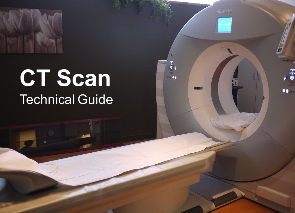
State-of-the-art CT scanner ready to transform patient care with high-resolution imaging.
This guide is brought to you by DataRay - The leaders in laser beam profilers.
1. Introduction
The advancement of Computed Tomography (CT) scan technology marks a significant milestone in medical diagnostics, offering multi-dimensional views into the human body with exceptional clarity. Its integration into routine healthcare underscores a revolution in detecting and treating diseases, with rapid imaging and detailed internal views facilitating swift and informed medical decisions. This continuous evolution in CT capabilities echoes its critical role in modern medical imaging, shaping patient outcomes and the future of radiological sciences.
Beyond diagnostics, CT technology has broadened its application spectrum, venturing into complex surgical planning, cancer treatment monitoring, and intricate vascular studies. The advent of high-speed, multi-slice CT scanners has drastically reduced scan times, enhancing patient comfort and expanding the utility of CT scans in emergency medicine. As we stand on the brink of integrating artificial intelligence with CT imaging, the potential for early disease detection, personalized treatment plans, and predictive analysis promises to redefine the standards of medical care and imaging precision.
2. Principles of CT Imaging
CT imaging, at its core, revolves around the sophisticated interplay of X-rays and computer processing to generate cross-sectional images of the body. This section delves into the foundational physics of X-ray generation and detection, the methodical process by which CT scanners construct detailed images, and the distinct characteristics that set CT apart from other imaging modalities like MRI, traditional X-ray, and ultrasound.
Physics Behind X-ray Generation and Detection
The generation of X-rays in a CT scanner begins with an electron beam directed at a tungsten target within the X-ray tube. The collision of high-speed electrons with the target material produces X-rays, which are then focused and directed toward the patient. As these X-rays pass through the body, they are attenuated to varying degrees by different tissues, based on their densities. Detectors positioned opposite the X-ray source capture the attenuated X-rays, converting the energy into electrical signals that reflect the internal structure of the scanned area.

Inside the CT scanner: The critical moment where high-speed electrons collide with tungsten, giving birth to the X-rays that illuminate the mysteries of the human body. Image courtesy of SchoolPhysics.
How CT Scanners Create Cross-sectional Images
CT scanners employ a rotating gantry that houses the X-ray source and detectors, circling around the patient to capture multiple projections from various angles. These projections are then reconstructed into cross-sectional images, or slices, by advanced computer algorithms. Unlike conventional X-rays that overlay all tissue densities onto a single image, CT scans differentiate between tissues in a slice, providing a detailed, slice-by-slice view of the body’s internal structures.
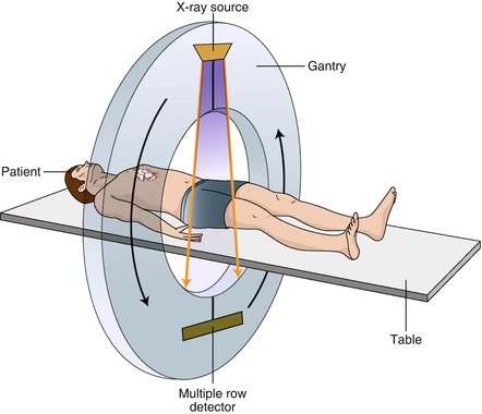
Journey through the layers: A glimpse into how CT scanners dissect complex anatomy into clear, cross-sectional slices, offering a window into the body’s hidden details. Image courtesy of Richard Garnett.
Differences Between CT and Other Imaging Technologies
While traditional X-rays offer quick and simple imaging solutions, they lack the depth and detail of CT scans, which provide comprehensive views of soft tissues, blood vessels, and bones with superior clarity. MRI (Magnetic Resonance Imaging), on the other hand, excels in soft tissue contrast without using ionizing radiation, making it preferable for certain types of examinations. However, MRI scans are more time-consuming and can be more challenging for claustrophobic patients. Ultrasound imaging uses sound waves to produce images, offering real-time, radiation-free examinations, particularly useful in obstetrics and cardiology. However, it is limited by its inability to penetrate bone and visualize deeper structures as clearly as CT or MRI.
To sum up, CT imaging harnesses the nuanced physics of X-ray generation and sophisticated computational algorithms to render detailed cross-sectional views of the body, setting it apart from other imaging modalities through its speed, precision, and versatility.
3. Components of a CT Scanner
The efficacy of Computed Tomography (CT) scanning is a result of its complex components working in harmony. In this section we examine the critical hardware elements—gantry, X-ray tube, detectors, and patient table—that constitute the CT scanner, as well as the sophisticated software responsible for image reconstruction, processing, and data storage.
Hardware Components
Gantry: The gantry is the framework that houses the key components of a CT scanner. It is characterized by a large, circular opening where the patient is positioned for scanning. Within the gantry, the X-ray tube and detectors are mounted on opposite sides and rotate around the patient to capture comprehensive imaging data from various angles.
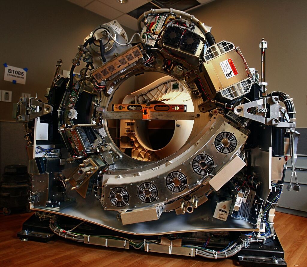
Behold the gantry: The heart of the CT scanner, where technology meets human care, transforming X-rays into clear visions of our internal health. Photo courtesy of Computed Tomography.
X-ray Tube: The X-ray tube is the heart of the CT scanner, responsible for generating X-rays. When activated, electrons within the tube are accelerated towards a tungsten target, producing X-rays that are then directed through the patient’s body. The design and efficiency of the X-ray tube are pivotal in defining the quality and speed of the scan.
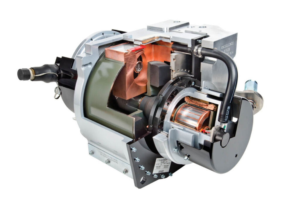
Inside the X-ray tube: The origin of diagnostic clarity, where electrons and tungsten converge to light up the unseen details of the human body. Image courtesy of 24×7.
Detectors: Positioned directly across from the X-ray tube, the detectors play a crucial role in capturing the X-rays that pass through the patient. These detectors convert the X-ray photons into electrical signals, which are proportional to the intensity of the X-rays received, thereby creating a digital map of the scanned area.
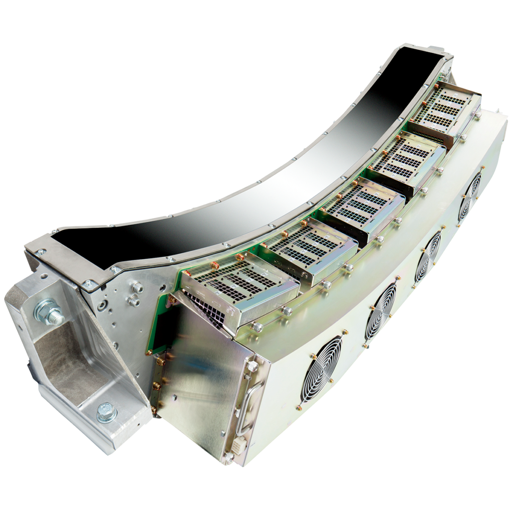
Detectors in action: The silent witnesses of the CT scanning process, translating X-rays into the digital blueprints of our internal landscape. Image courtesy of Canon.
Patient Table: The patient table is designed for comfort and precision, able to move smoothly through the gantry during the scan. Its movement is meticulously synchronized with the gantry’s rotation, ensuring that images are captured at the correct positions and angles for accurate reconstruction.
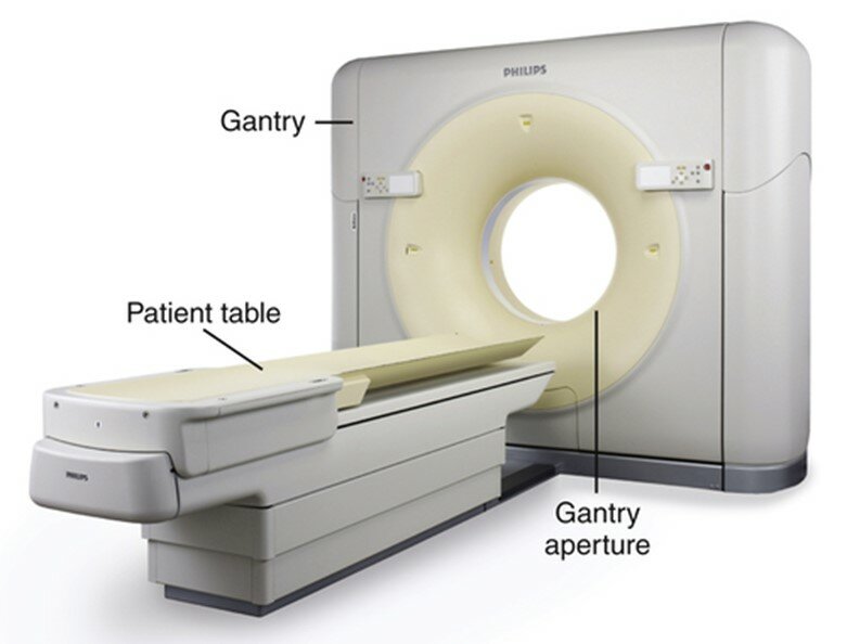
The patient table: A blend of precision engineering and patient comfort, ensuring every scan captures the perfect angle for revealing insights. Image courtesy of ResearchGate.
Software Components
- Reconstruction Algorithms: The core of CT image processing, these algorithms transform the raw data collected by the detectors into interpretable images. Techniques like filtered back projection and iterative reconstruction are used to compile the data from various angles into coherent cross-sectional images, providing the detail and clarity that CT is known for.
- Image Processing: Beyond initial reconstruction, additional image processing is applied to enhance image quality. This includes adjustments for contrast, noise reduction, and edge sharpening, which help in improving the diagnostic utility of the images.
- Data Storage Solutions: Managing the vast amount of data generated by CT scans requires robust data storage solutions. Modern CT systems are integrated with PACS (Picture Archiving and Communication Systems) and cloud-based storage, facilitating efficient data management, retrieval, and sharing within medical networks.
The integration of advanced hardware with innovative software algorithms underpins the CT scanner’s ability to deliver rapid, high-resolution imaging, making it a cornerstone of contemporary diagnostic imaging.
4. CT Imaging Techniques
CT imaging techniques have evolved significantly, offering a range of methodologies tailored to various diagnostic needs. In this section we look into three key techniques: Spiral (Helical) CT scanning, Multi-Detector CT (MDCT), and Dual-Energy CT scanning, each enhancing the scope and precision of CT imaging.
Spiral (Helical) CT Scanning
Spiral, or Helical, CT scanning represents a leap from traditional axial scanning by continuously rotating the X-ray source and detectors around the patient as the table moves through the gantry. This motion traces a helical path, enabling the acquisition of data in a single, smooth motion, significantly reducing scan times and improving image quality. The continuous data stream facilitates the reconstruction of images at any point along the scanned volume, offering greater flexibility in image review and interpretation.
Multi-Detector CT (MDCT)
The advent of Multi-Detector CT (MDCT) systems, equipped with multiple rows of detectors, has exponentially increased data acquisition speed and image resolution. MDCT scanners can capture multiple slices simultaneously, dramatically shortening scan times and enhancing image detail. This capability is particularly beneficial for cardiac, trauma, and pediatric imaging, where motion artifacts can be minimized, and sedation times reduced. The increased speed and resolution of MDCT also improve the quality of 3D reconstructions, aiding in complex diagnostic and surgical planning.
Dual-Energy CT Scanning
Dual-Energy CT scanning utilizes two different X-ray energy levels to differentiate materials within the body based on their atomic number. By capturing images at both high and low energies, this technique can distinguish between substances with similar densities but different chemical compositions, such as distinguishing between iodine-containing contrast material and calcium in blood vessels. Applications of Dual-Energy CT include improved detection and characterization of lesions, virtual non-contrast imaging, and advanced applications in oncology and vascular imaging. The ability to extract material-specific information from CT data opens new avenues for non-invasive diagnostics and precise disease characterization.
These advanced CT imaging techniques enhance the diagnostic capabilities of CT scans, enabling more detailed and specific evaluations of anatomical structures and pathologies. The continuous innovation in imaging techniques underscores the versatility and adaptability of CT technology in meeting diverse clinical demands.
5. Image Reconstruction and Enhancement
The transformation of raw CT data into clinically useful images is a sophisticated process involving advanced reconstruction algorithms and enhancement techniques. Let’s explore the key reconstruction algorithms like Filtered Back Projection and Iterative Reconstruction, outlines methods for noise reduction and image quality improvement. Here was also touch on the emerging role of artificial intelligence in image reconstruction.
Reconstruction Algorithms
- Filtered Back Projection (FBP): Traditionally the backbone of CT image reconstruction, FBP algorithms compile the raw data collected from multiple angles into a coherent image. This method applies a filter to the raw data to counteract the blurring effect inherent in the back-projection technique. While FBP is fast and computationally efficient, it can be susceptible to noise, especially in low-dose CT scans.
- Iterative Reconstruction (IR): A more advanced approach, Iterative Reconstruction, progressively refines the image by comparing the simulated projections of an initial image estimate to the actual acquired data, making adjustments to minimize discrepancies. This method is particularly effective in reducing noise and improving image quality, making it invaluable for low-dose CT applications. The iterative process, although computationally intensive, offers superior image clarity and detail, with significant reductions in artifacts.
Techniques for Noise Reduction and Image Quality Improvement
Image quality in CT scanning can be compromised by factors such as patient movement, low signal-to-noise ratio, and artifacts. Techniques to mitigate these issues include:
- Beam Hardening Correction: Adjustments made to counteract the effects of beam hardening, where lower-energy photons are absorbed more readily than higher-energy ones, leading to artifacts in the reconstructed image.
- Adaptive Filtering: These filters adjust to the varying levels of noise across an image, smoothing out areas with high noise while preserving the integrity of detailed structures.
- Automatic Exposure Control (AEC): AEC systems adjust the radiation dose based on the part of the body being scanned, optimizing the balance between image quality and patient radiation exposure. You can read more about it here.
The Role of Artificial Intelligence in Image Reconstruction
Artificial Intelligence (AI) is revolutionizing CT image reconstruction by leveraging deep learning algorithms to enhance image quality and reduce artifacts without increasing radiation dose. AI-driven methods can predict optimal reconstruction parameters, identify and reduce artifacts, and improve the signal-to-noise ratio, thereby enhancing the diagnostic utility of CT images. The integration of AI into image reconstruction workflows promises not only to improve image quality but also to streamline the imaging process, making it faster and more efficient.
Through these sophisticated reconstruction and enhancement techniques, CT imaging continues to advance, offering clearer, more detailed images that enhance diagnostic accuracy and patient care.
6. Advanced Applications of CT Technology
CT technology has evolved beyond simple diagnostic imaging, embracing advanced applications that extend its utility into 3D modeling, vascular studies, and quantitative analysis. This section highlights the sophisticated use of 3D CT imaging in surgical planning, the critical role of CT angiography and perfusion studies in assessing vascular health, and the application of Quantitative CT (QCT) in evaluating bone density.
3D CT Imaging and Its Applications in Surgery and Treatment Planning
3D CT imaging transforms traditional two-dimensional slices into three-dimensional models, offering a comprehensive view of anatomical structures. This innovation is particularly impactful in the realm of surgery and treatment planning, where precise anatomical details are crucial. Surgeons utilize 3D models for preoperative planning, simulating surgical procedures to determine the most effective approaches. In complex cases, such as reconstructive surgery or tumor removal, 3D CT imaging enables a level of precision that significantly enhances surgical outcomes and patient safety.
CT Scan Angiography and Perfusion Studies
CT angiography (CTA) employs contrast material to visualize blood vessels, providing detailed images that assist in diagnosing vascular diseases, such as aneurysms and blockages. The non-invasive nature of CTA makes it a valuable tool for quickly assessing patients with symptoms of cardiovascular disease. Perfusion CT, on the other hand, measures blood flow to various tissues, crucial in stroke management where identifying viable brain tissue can dictate treatment decisions. These techniques exemplify how CT technology contributes to dynamic vascular assessment, offering real-time insights into blood flow and vessel integrity.
Quantitative CT (QCT) for Bone Density Analysis
QCT specializes in measuring bone mineral density (BMD), offering a valuable tool in diagnosing and monitoring osteoporosis. Unlike traditional DEXA scans, QCT provides a three-dimensional assessment of bone density, allowing for precise measurements of the spine and other skeletal sites. This quantitative approach offers detailed insights into bone health, enabling personalized treatment plans to combat osteoporosis and assess fracture risk more accurately. QCT’s ability to provide volumetric density measurements highlights the adaptability of CT technology in addressing specific medical concerns.
These advanced applications of CT technology not only broaden its diagnostic capabilities but also introduce new dimensions to patient care, from pre-surgical planning to targeted treatment strategies, showcasing the versatility and continuous innovation within CT imaging.
7. Innovation in CT Scan Design and Functionality
The landscape of CT technology is constantly evolving, with innovations aimed at enhancing healthcare delivery, patient safety, and comfort. This section explores the advent of portable CT scanners, advancements in patient-centric design, and the concerted efforts to reduce radiation exposure, underscoring the dynamic nature of CT scan design and functionality.
Portable CT Scanners and Their Impact on Healthcare Delivery
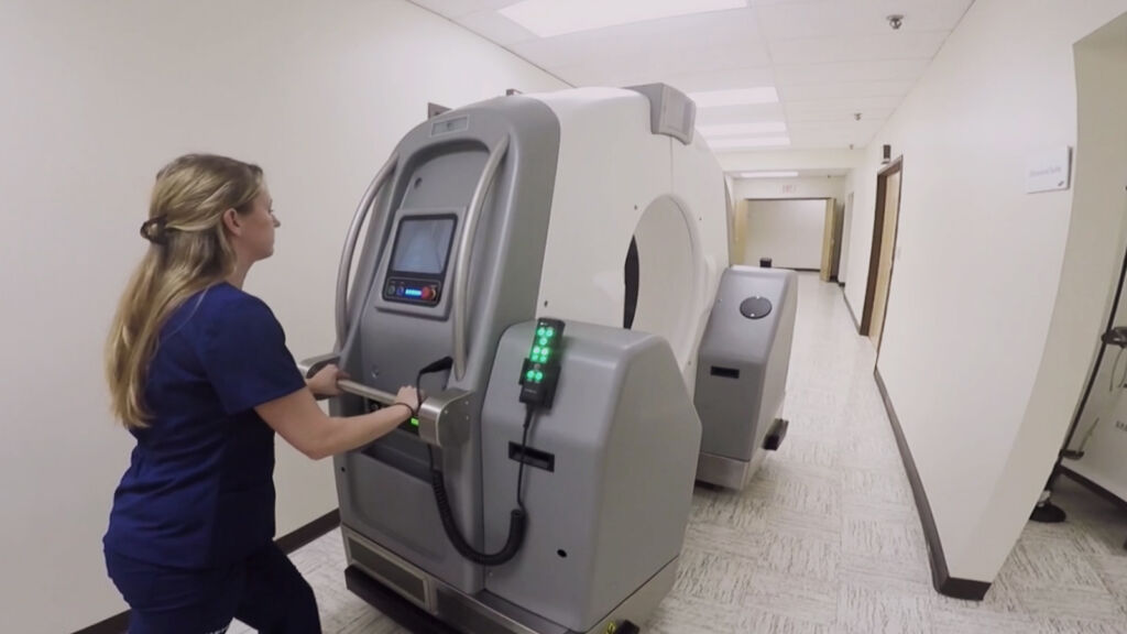
Bringing technology to the patient: A nurse navigates a portable CT scanner through the hospital, showcasing the fusion of mobility and cutting-edge medical imaging. Photo courtesy of Samsung Health.
The development of portable CT scanners marks a significant leap forward in medical imaging, bringing diagnostic capabilities directly to the patient’s bedside. This mobility is particularly transformative in critical care units, emergency departments, and remote locations where traditional CT infrastructure is inaccessible. Portable CT scanners expedite diagnosis and treatment decisions for conditions like strokes or head injuries, dramatically improving patient outcomes by reducing the time to intervention. Their integration into healthcare systems exemplifies how technology can adapt to meet clinical needs, enhancing accessibility and responsiveness in patient care.
Advances in Patient Safety and Comfort
Modern CT scanners are designed with a strong emphasis on patient safety and comfort. Innovations include wider gantries and shorter bore lengths to alleviate claustrophobia, noise reduction technology for a more pleasant scanning experience, and faster scanning speeds to minimize discomfort and the need for prolonged stillness. Enhanced patient positioning aids and ambient features, such as lighting and visual projections, further contribute to a less intimidating and more comfortable scanning environment. These patient-centric designs are crucial in ensuring that the CT scanning process is as stress-free as possible, encouraging patient compliance and facilitating optimal imaging outcomes.
Reduction of Radiation Dose: Techniques and Strategies
Minimizing radiation exposure while maintaining image quality is a central focus in CT scan innovation. Techniques such as iterative reconstruction algorithms allow for lower radiation doses by improving the efficiency of image processing, resulting in high-quality images from less radiation. Automatic exposure control systems adjust the dose based on the specific area being scanned, optimizing the balance between image clarity and radiation safety. Developments in detector technology also contribute to dose reduction, with more sensitive detectors requiring fewer X-rays to produce clear images. These strategies exemplify the commitment to “As Low As Reasonably Achievable” (ALARA) principles in CT imaging, ensuring patient safety without compromising diagnostic precision.
Through these innovations in design and functionality, CT technology continues to advance, prioritizing patient care, safety, and the ever-important reduction of radiation exposure, all while enhancing the diagnostic capabilities that make CT an essential tool in modern medicine.
8. CT Scan Data: Management and Analysis
The management and analysis of CT imaging data are critical components of modern medical imaging, encompassing the acquisition, storage, and sophisticated handling of vast amounts of data. This section delves into the efficient management of CT data, the role of advanced visualization software in interpreting complex images, and the seamless integration of CT systems with broader hospital information infrastructures.
Data Acquisition, Storage, and Management in CT Scan
CT scans generate large volumes of data, with multi-detector systems producing thousands of images per scan. Efficient data acquisition systems are essential, ensuring that images are captured, processed, and ready for review swiftly and accurately. Once acquired, the data must be securely stored and managed, often requiring substantial digital storage solutions. Data management protocols ensure the integrity and confidentiality of patient information, employing sophisticated data compression and encryption technologies to optimize storage efficiency and safeguard patient privacy.
Advanced Visualization Software for CT Scan Data
The complexity of CT data necessitates advanced visualization software, capable of rendering multi-dimensional images for detailed analysis. This software allows radiologists and medical professionals to navigate through layers of images, adjust parameters for optimal viewing, and apply algorithms that can highlight specific structures or pathologies. 3D reconstruction and volume rendering capabilities transform traditional slices into intuitive, spatial representations of anatomy, facilitating a deeper understanding of patient conditions and enhancing diagnostic accuracy.
Integration with Hospital Information Systems and PACS
The integration of CT imaging systems with Hospital Information Systems (HIS) and Picture Archiving and Communication Systems (PACS) is crucial for the streamlined operation of modern healthcare facilities. This integration enables the efficient transfer, retrieval, and sharing of imaging data within a secure network, ensuring that patient information is accessible to authorized professionals across departments. The compatibility with Electronic Health Records (EHR) systems further enhances clinical workflows, allowing for a comprehensive view of patient health records, including CT images, directly within the patient’s file. This interconnectedness supports multidisciplinary collaboration, informed decision-making, and continuity of care, exemplifying the pivotal role of IT infrastructure in optimizing the use and impact of CT imaging in healthcare.
Through meticulous data management, advanced visualization tools, and seamless integration with hospital systems, CT imaging continues to be at the forefront of diagnostic medicine, offering unparalleled insights while ensuring efficiency and security in handling patient data.
9. Quality Control and Maintenance
Ensuring the consistent quality of CT images and the optimal functioning of CT scanners is crucial in diagnostic imaging. This section outlines the rigorous protocols for quality control, the importance of routine maintenance, and the critical role of technologists in maintaining quality assurance in CT imaging.
Protocols for Ensuring and Maintaining the Quality of CT Images
Quality control protocols in CT imaging are comprehensive, designed to maintain the accuracy, safety, and reliability of diagnostic images. These protocols encompass regular calibration of the CT scanner to ensure precise imaging parameters, phantom studies to assess image quality and scanner performance, and adherence to standardized imaging protocols to minimize variability. Regular audits of image quality, including resolution, contrast, and noise levels, are conducted to identify and rectify any deviations from established benchmarks.
Routine Maintenance of CT Scanners
Routine maintenance of CT scanners is essential to prevent downtime and extend the equipment’s lifespan. Preventive maintenance schedules are strictly followed, involving the inspection and servicing of key components such as the X-ray tube, gantry, and detectors. Software updates and hardware upgrades are also part of routine maintenance, ensuring that the CT system operates with the latest advancements in imaging technology. Emergency repair protocols are in place to address unexpected issues, minimizing disruptions to diagnostic services.
The Role of the Technologist in Quality Assurance
Technologists play a pivotal role in quality assurance within CT imaging. Their expertise in operating CT scanners, executing imaging protocols, and managing patient positioning directly influences the quality of the images produced. Technologists are also the first line of defense in identifying image anomalies or equipment malfunctions, initiating corrective actions, and liaising with maintenance teams. Continuous professional development keeps technologists abreast of best practices and technological advancements, reinforcing their essential contribution to maintaining high standards in CT imaging quality and patient care.
Through diligent adherence to quality control protocols, meticulous maintenance routines, and the dedicated involvement of technologists, the integrity and reliability of CT imaging are upheld, ensuring its continued efficacy in diagnostic medicine.
10. Future of CT Scan Technology
The horizon of CT scan technology is marked by rapid advancements and promising research that aim to redefine imaging capabilities. In this section we explore the emerging technologies poised to enhance CT imaging, delves into the cutting-edge research areas like nanotechnology and photon-counting detectors, and offers predictions for the next generation of CT scanners.
Emerging Technologies in CT Scan Imaging
Innovation in CT technology is focused on improving image quality, reducing scan times, and minimizing radiation exposure. Developments such as spectral (or dual-energy) CT provide enhanced material differentiation, offering new insights into tissue composition and function. Artificial intelligence and machine learning algorithms are being integrated to optimize scan parameters in real-time, improve image reconstruction, and assist in automated lesion detection and diagnosis, promising a future where CT imaging is faster, more accurate, and safer.
Research Frontiers: Nanotechnology, Photon-Counting Detectors
Nanotechnology in CT imaging introduces nanoparticles as contrast agents, potentially enabling highly targeted imaging of cellular and molecular processes, opening new avenues in early disease detection and therapy monitoring. Photon-counting detectors represent another frontier, offering superior resolution and contrast by counting individual photons and measuring their energy. This technology promises significant improvements in image quality and a substantial reduction in radiation doses, paving the way for safer and more precise diagnostic imaging.
Predictions for the Next Generation of CT Scan Machines
The next generation of CT scanners is anticipated to be characterized by enhanced integration with other imaging modalities, such as PET or MRI, providing comprehensive diagnostic information in a single session. Advances in hardware, such as more efficient X-ray tubes and detector materials, along with software innovations, are expected to deliver higher resolution images at lower doses. Portability and accessibility of CT technology are likely to expand, making advanced imaging available in a wider range of clinical settings, including remote and underserved areas.
The future of CT technology holds the promise of transformative changes, with ongoing research and development focused on pushing the boundaries of what is possible in medical imaging, enhancing the role of CT in patient care, and opening new vistas in medical research and diagnostics.
11. Understanding CT Scan Technology for Engineers
For developers and engineers, CT scan technology offers a dynamic landscape for innovation and collaboration. In this section we shed light on the essential technical skills needed to drive advancements in CT imaging, highlights the opportunities for innovation, and emphasizes the importance of interdisciplinary collaboration in pushing the boundaries of what’s possible in medical imaging.
Technical Skills Required for Developing CT Scan Technology
Developing CT technology demands a robust foundation in physics, particularly in understanding X-ray generation and detection, as well as expertise in electrical and mechanical engineering to design and optimize the hardware components of CT scanners. Proficiency in computer science is crucial for developing advanced image reconstruction algorithms, data processing techniques, and integrating AI into CT imaging. Familiarity with biomedical engineering principles also plays a significant role, ensuring that technological advancements align with clinical needs and safety standards.
Opportunities for Innovation in CT Imaging
The field of CT imaging is ripe with opportunities for innovation, ranging from enhancing image quality and reducing scan times to minimizing radiation exposure. Engineers and developers can explore advancements in detector technology, such as developing more sensitive materials that require less radiation. Innovations in software, including machine learning algorithms for image analysis and reconstruction, present another avenue for enhancing the capabilities of CT imaging. There’s also potential in exploring novel contrast agents and imaging techniques that provide new types of diagnostic information, expanding the clinical applications of CT scans.
Collaboration Between Engineers, Technologists, and Clinicians
The development of effective and impactful CT technology relies heavily on the collaboration between engineers, technologists, and clinicians. Engineers and developers bring technical expertise, while technologists offer insights into the practical aspects of CT imaging, including operational efficiencies and patient interaction. Clinicians provide the clinical context, identifying unmet needs and guiding the development of technologies that address real-world challenges. This interdisciplinary approach ensures that innovations in CT imaging are not only technologically advanced but also clinically relevant and focused on improving patient care.
Understanding CT scan technology from a development and engineering perspective underscores the importance of technical proficiency, creative innovation, and collaborative efforts in advancing medical imaging. It’s through this synergy that CT technology will continue to evolve, enhancing diagnostic capabilities and ultimately benefiting patient outcomes.
12. Resources for Technologists and Biomedical Engineers
Staying at the forefront of CT technology requires continuous learning and engagement with the professional community. Here we outline a few valuable resources for professional development, including leading journals and conferences in medical imaging technology, as well as online platforms and communities that serve as hubs for knowledge exchange and networking for CT technologists and biomedical engineers.
Professional Development in CT Scan Technology
Technologists and engineers looking to excel in CT imaging can benefit greatly from targeted training programs and certifications offered by recognized institutions and professional bodies. For instance, the American Registry of Radiologic Technologists (ARRT) offers certification in CT, which is widely recognized and often required by employers. Similarly, the American Society of Radiologic Technologists (ASRT) provides a wealth of continuing education (CE) opportunities tailored to CT technology.
Specialized courses often cover a wide array of topics, from foundational principles of CT imaging and patient safety protocols to intricate details of advanced imaging techniques and the latest technological innovations. For example, programs may include in-depth training on the use of dual-energy CT applications, mastery of 3D reconstruction software, and strategies for minimizing radiation exposure while maximizing image quality.
Professionals seeking to push the boundaries of their expertise might also explore advanced workshops and seminars focused on emerging areas like artificial intelligence applications in CT imaging, offered by academic institutions or through industry-led training platforms like Siemens Healthineers, GE Healthcare, or Philips Learning Center. These courses not only provide practical knowledge and skills but also offer valuable CEUs that contribute to the maintenance of certification and professional growth.
Engaging in these educational pathways not only sharpens current skills but also opens doors to new opportunities in the field of CT imaging, positioning technologists and engineers at the forefront of medical imaging innovation.
Leading Journals and Conferences in Medical Imaging Technology
Engaging with the latest research and developments in medical imaging is essential for professionals in the field. Leading journals such as “Radiology,” “Journal of Medical Imaging,” and “Medical Physics” publish peer-reviewed articles on cutting-edge CT technology and applications. Conferences like the Radiological Society of North America (RSNA) Annual Meeting, the European Congress of Radiology (ECR), and the International Society for Computed Tomography (ISCT) conference provide platforms for presenting research, attending workshops, and networking with peers and industry leaders.
Online Platforms and Communities for CT Scan Technologists and Engineers
Online platforms and professional communities offer invaluable resources for sharing knowledge, discussing challenges, and staying updated on industry trends. Websites like AuntMinnie.com and Radiopaedia.org serve as comprehensive repositories for radiology education and case studies. Social media groups, forums, and LinkedIn communities dedicated to CT technology foster discussions on technical issues, career advice, and innovations in the field. Engaging with these online communities can provide support, inspiration, and opportunities for collaboration.
For technologists and biomedical engineers in the field of CT imaging, accessing these resources can significantly contribute to professional growth, keeping them informed about the latest technological advancements and best practices in medical imaging.
13. Conclusion
The journey through the technological landscape of CT scanning reveals a field rich with innovation, precision, and transformative potential in healthcare. From its inception to the cutting-edge advancements of today, CT technology has continually redefined the boundaries of medical imaging, offering deeper insights into the human body with unprecedented clarity and efficiency.
The pivotal role of CT scanning in advancing healthcare cannot be overstated. It has revolutionized diagnostic practices, enabling rapid, non-invasive detection and assessment of a wide range of conditions, thus facilitating timely and targeted treatment interventions. The evolution of CT technology reflects a broader commitment within the medical community to enhance patient care, improve outcomes, and extend the horizons of medical research.
As we stand on the threshold of future innovations, the importance of ongoing education and engagement with the latest developments in the field becomes ever more critical. For technologists, engineers, and clinicians alike, embracing continuous learning and contributing to the innovation ecosystem will ensure that CT technology remains at the forefront of medical imaging. The collective endeavor towards refining and expanding the capabilities of CT scanning promises not only to advance the field of radiology but also to open new avenues for patient care and treatment possibilities.
In conclusion, the technological marvel of CT scanning is a testament to the ingenuity and dedication of the medical and scientific communities. As we look forward, let us continue to champion education, innovation, and collaboration, driving forward the capabilities of CT technology to meet the evolving demands of healthcare and to forge new paths in the quest for excellence in patient care.
