Dental caries, often referred to as tooth decay or cavities, is one of the most common oral diseases worldwide. It affects the majority of children, as well as large percentage of adults. In the US, there are more than 3 million cases per year. Early diagnosis is important for assessment and treatment, before the disease can progress. Image courtesy of Pixabay.
A research team from the Military Technical College and from Cairo University in Cairo, Egypt have developed a fluorescence hyper-spectral imaging system which can be employed for diagnosis and detection of dental caries without causing mechanical or thermal harm to the teeth being inspected. A paper recently published by the team describes the system and its results.
What are dental caries?
Human tooth enamel, dentin, and cementum, three components of teeth, are classified as hard tissue, or calcified tissue. The mineralized tissue of the enamel coats the tooth, acting as a barrier and protecting it. However, it is susceptible to degradation, often due to acid present after the consumption of sugary foods and drinks. When an individual eats something containing lots of sugar, the sugar reacts with bacteria present in the plaque on the teeth. This, in turn, produces acid, which can demineralize the tooth by degrading the calcium and phosphate in the enamel.
Over time, this demineralization leads to dental caries, commonly known as cavities. Even when such cavities exist in the non-permanent teeth of children, they cause extreme pain and can lead to infections and chewing problems. To treat a common cavity, the decay is drilled out and a filling is put in. A larger cavity may require a crown, a root canal treatment, or extraction of the tooth. Therefore, to prevent further pain and extensive dental procedures, early detection is crucial. Current detection techniques include visual and tactile inspections, which lack specificity and sensitivity, and radiography, which poses a radiation threat to patients.
How does the imaging system work?
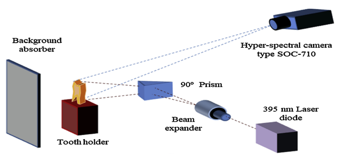
The goal of hyper-spectral imaging is measuring the electromagnetic spectrum relevant to each pixel of a scene in order to identify specific objects, materials, or processes. The hyper-spectral imaging system, as shown below, consists of a laser diode source, a beam expander, and a hyper-spectral camera, along with a few other accessories. Associated software processes the raw data from the hyper-spectral camera, and displays the reconstructed data.

The diagram shown above displays the experimental set-up for the fluorescence hyper-spectral imaging system. Image courtesy of Photodiagnosis and Photodynamic Therapy 25.
The imaging system is based on laser-induced fluorescence. This phenomenon sees an interaction between the irradiating wavelength- 395nm in this experiment- with the fluorophores present in the hard tissues of the tooth sample being irradiated by the laser. When the fluorophores are excited due to irradiation, they emit a higher wavelength. The bacterial components found in the dental caries also contain fluorophores, which exhibit laser-induced fluorescence properties as well. The camera, which uses advanced fluorescence spectroscopic and hyper-spectral imaging techniques, detects and measures this activity. Researchers compared the processed data with preliminary knowledge of each tooth sample regarding its state and locations of decay, obtained through visual and tactile observations.
Results
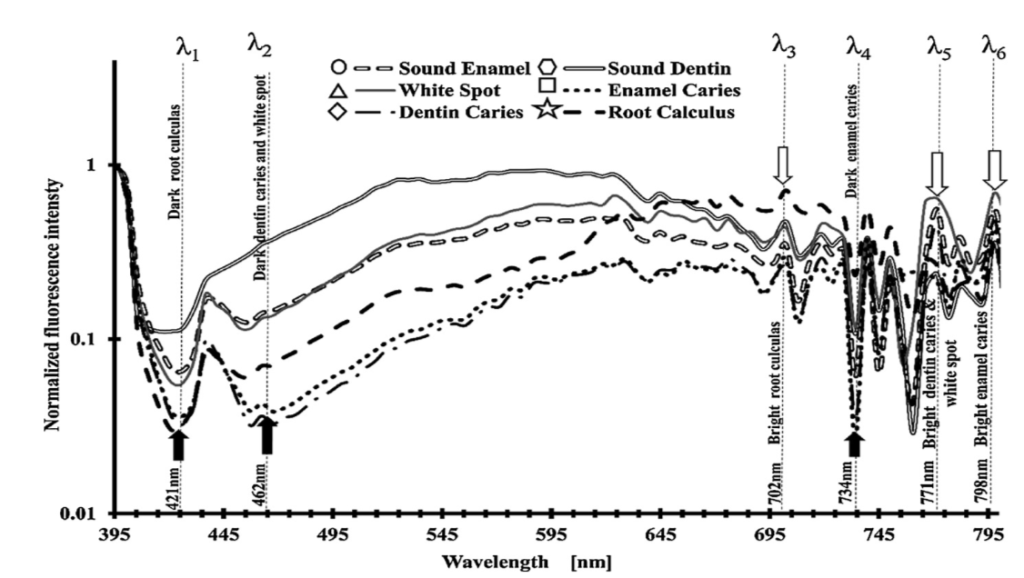
Researchers focused on the one-dimensional spectroscopic measurements as well as the grayscale two dimensional images obtained at deduced wavelengths. The one-dimensional spectroscopic measurements had to be normalized to enhance differences between the various dental diseases.

The figure above displays the processed fluorescence data of dental classes, showing lower and higher peak wavelengths. This data was used to select the six wavelengths used for later analysis. Image courtesy of Photodiagnosis and Photodynamic Therapy 25.
Wavelengths corresponding to the high and low peaks of the normalized fluorescence graph were identified. At these wavelengths, the various oral diseases would appear as either dark or bright.

The table above displays the contrast visible for multiple dental diseases as a function of the obtained LIF wavelengths. Image courtesy of Photodiagnosis and Photodynamic Therapy 25.
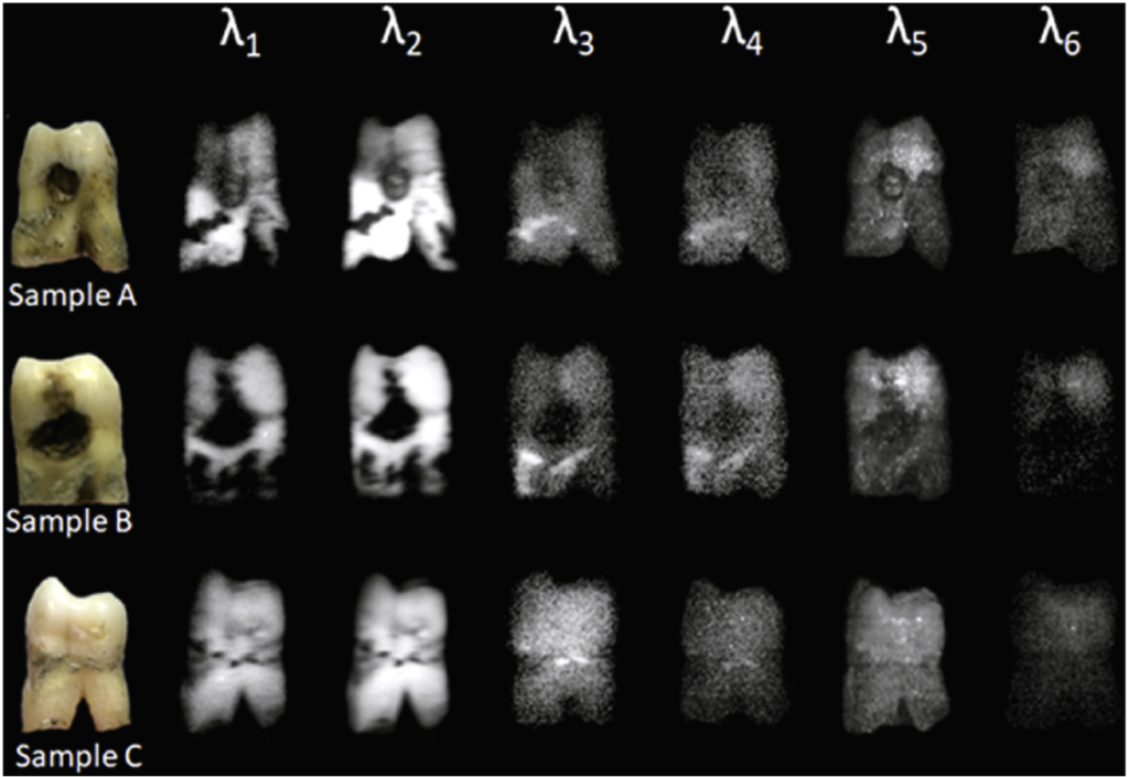
These wavelengths obtained were then used further for the analysis of the two-dimensional data. By applying these obtained wavelengths, an image of each tooth sample could be constructed displaying light and bright spots corresponding to the dental diseases. For example, in the figure below, at λ2, where dental caries are shown as dark spots, it is clear that both Sample A and Sample B have dental caries. This was confirmed using the initial tactile and visual observations.

The figure above displays the two-dimensional hyper-spectral images obtained for three tooth samples at the six determined wavelengths. Image courtesy of Photodiagnosis and Photodynamic Therapy 25.
The wavelengths used were based on the average of a very small sample size. Therefore, further experimentation and analysis of samples would be necessary to obtain a better set of wavelengths for more specific and sensitive data analysis and detection of dental caries.
Conclusion
This new imaging system was capable of producing tooth auto-fluorescence. The hyper-spectral camera could collect and, along with associated software, successfully reconstruct the fluorescence emissions. Researchers discovered sets of estimated wavelengths corresponding to several dental abnormalities. Future fine-tuning of the device to improve quality and quantification of oral diseases is necessary before it can be used by dentists and oral surgeons. Additionally, the effect of the device on teeth should be studied as well. However, in conclusion, this fluorescence hyper-spectral oral imaging device shows great potential to be a low-cost, easy to use, non-invasive detection device for professionals in the dental field. It could also potentially have future uses in other medical fields, regarding other hard calcified tissues such as bone.
To read more about this dental imaging system, read the paper here.

Thank You for sharing such a nice and informative blog and your knowledge with us. Tooth decay, also known as dental caries or cavities, is a breakdown of teeth due to acids made by bacteria. The cavities may be a number of different colors from yellow to black. Symptoms may include pain and difficulty with eating.