Since the invention of the first operating laser in 1960, lasers have been designed in various regions of the electromagnetic spectrum and output powers. Soon after the advent of semiconductor fabrication techniques in the late 20th century, technology evolved to give birth to the first diode lasers using semiconductors. Over the ensuing decades, the use of such lasers in combination with advanced electronics enabled paradigm changing applications from telecommunications to medicine.
Today, diode lasers differ from their tabletop counterparts by being smaller in size, and delivering smaller output powers. They also differ from fiber lasers, and solid-state lasers in the way optical amplification occurs, yielding rather different output characteristics. For example, optical pumping is done by current injecting in laser diodes. Miniaturization helps increase throughput in producing mass quantities of diode lasers, as is the case in the manufacturing of VCSELs. With the constant need to make devices smaller, laser diode technology evolved to making quantum well lasers, and quantum dot lasers. We will not touch upon the elements of these specific lasers in this article, as we have talked about them and their applications in another FindLight blog post in some detail.
Here we look into the applications of laser diodes for cytometry. Cytometry is the analysis of cells to gain information about their structure and content. Recently, the use of laser diodes has become popular for this technique as one can measure the scattered light to learn a lot about cells, in particular whether they are cancerous or not. Moreover, this process has the potential for automation, if it can be integrated with an automatic detection and analysis circuitry.
What is Cytometry?
Cytometry is the quantitative analysis of cell’s properties. Cell properties such as the size, shape, and content can all be inferred from cytometry techniques. Currently there are two main flavors of this technique: image and flow cytometry. In image cytometry, optical microscopy is used to image the group of cells and infer information from image processing techniques. In flow cytometry, the cells are aligned and allowed to flow in a line one at a time to infer individual cell characteristics. Usually additional circuitry is needed to collect data analyzed from each cell for further processing. Flow cytometry has gained popularity over the past few years, as it has the benefit of not requiring image processing to infer properties. Single cell techniques such as this can be made easy using diode lasers, which we discuss in the following section of this blog post.
Cytometry has the potential to make crucial advances in the field of molecular biology. Information about the cell’s aging phase and content can help identify its cancerous nature. Moreover, high throughput processing techniques in biology can benefit from automated analysis of cell’s properties.
Diode Lasers for Cytometry
It turns out, lasers can be used for analyzing and sorting cells. To accomplish this, cells are first “stained” by injecting them with fluorophores – compounds that emit light in the visible region when hit with incoming light. Stained cells then become easy targets for optical differentiation and analysis.


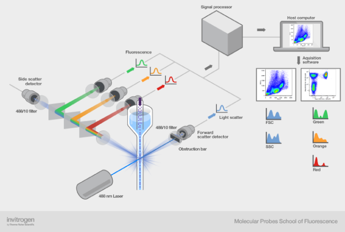
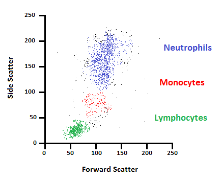
Top: Setup with the cell injection and laser to determine the properties. Bottom: Forward and side scattering for various types of cells at different wavelengths. Courtesy of ThermoFisher
A typical setup involves a collimating funnel which sends down cells to analyze from the top. The laser beam hits one cell at a time and the scattered light is detected using photodetectors and accompanying electronics to distinguish the type of cell. For the purpose of cell counting and detection, diode lasers in the visible region are used, as fluorophores operate in the visible region. There are two types of scattering that can occur from cells: forward and side scattering with varying intensities depending on the cell type. Moreover, when injected with fluorophore dye compounds, analysis is enhanced with reflection only happening at certain wavelengths. The 2D scatter plot above indicates the regions and wavelengths in the 2D region used to detect various cells based on this method.

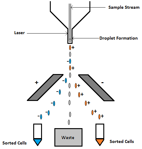
Cell sorting by lasers. Cells contain fluorophores which are activated by the lasers to charge the cells with a certain polarity. The cells are then separated into different polarity bins. Courtesy of Labome.
Conclusion
Cytometry is a powerful technique to obtain crucial information about the make up and nature of various cell structure. There is no need to emphasize the crucial role they play in modern medicine and the advances they foster in state of the art bio-engineering. The diagnostic power of this technique becomes especially formidable in combination with diode lasers. In particular, this method has been extended for other applications such as cell sorting in addition to counting and detection. Diode lasers have many interesting and far reaching applications because of miniaturization and capability to integrate with electronics.
Are you in the market for diode lasers? Check out FindLight’s collection of diode lasers from numerous world class suppliers to incorporate them into your project today.
