FFOCT could revolutionize corneal imaging, leading to accurate and immediate diagnosis to prevent blindness. Courtesy of WonderHowTo.
A new study presents a novel approach to imaging the human cornea. The research team at the Langevin Institute in Paris says their optical coherence tomography device is the first capable of full-field in vivo imaging of the cornea for preemptive diagnosis.
According to the Proctor World Blindness Center, between 1.5 and 2 million people go blind each year. Corneal disease is one of the leading causes of blindness, but early diagnosis allows for preventative treatment. It is therefore imperative to establish examination methods that produce real-time results. Accurately diagnosing the disease typically requires microscopic analysis because many macroscopic symptoms are indistinguishable from one another. Current methods in corneal imaging include both non-invasive and invasive techniques, both with glaring faults.

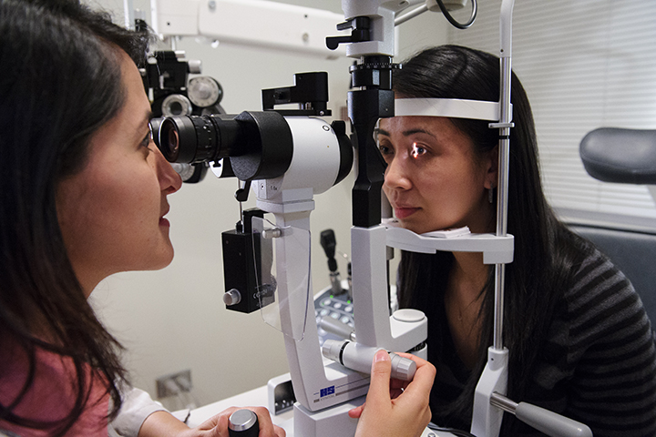
The slit lamp is the most common eye examination method. Courtesy of North Shore University.
What Are the Current Methods in Corneal Imaging?
The most well-known eye examination method is slit lamp biomicroscopy. Most glasses or contacts wearers should recognize the device. A thin sheet of light sweeps across the eye while a doctor peers through biomicroscope to look for irregularities. This method is reliable for a general exam, but it does not provide adequate resolution for an in depth examination of the cornea.
Another common method involves scraping the ocular surface for samples to be analyzed under microscope. Though this technique does offer higher resolution, it is very invasive and has a high false-negative rate.
Finally, confocal microscopy has become a much more useful option in the past few years. This method increases optical resolution of a micrograph by using a pinhole to block light that is out of focus. This offers high resolution, but axial and lateral resolutions are dependent on each other. This results in a limited field of view.

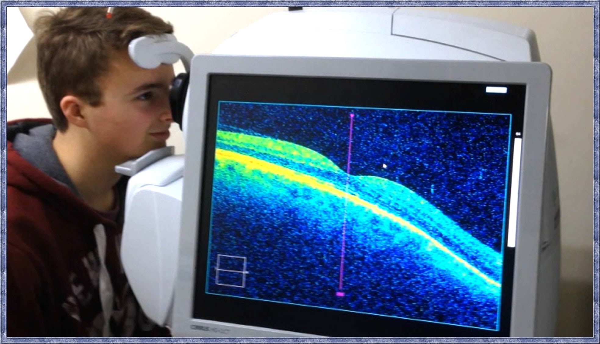
OCT offers high-resolution results in real time, but it suffers from a limited field of view. Courtesy of YouTube.
What Is Optical Coherence Tomography (OCT)?
In their innovative studies, the research team sought out to improve standard optical coherence tomography (OCT). OCT is an imaging technique that is capable of capturing micrometer-resolution using a coherent light source. The method employs near infrared light to create an interference pattern of the cornea. OCT is non-invasive and provides a high resolution image. Current OCT technology, however, suffers from a limited field of view and only provides cross-sectional images.
The Langevin research team thereby proposed a device capable of full-field optical coherence topography (FFOCT). They describe their solution as combining “the penetration capability and high axial resolution section of OCT with the high transverse resolution of confocal microscopy.” They effectively decouple the axial and transverse resolutions to enable a larger field of view. The final product acquires two-dimensional en face optical slices that are directly recorded to a camera.

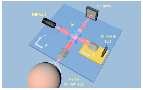
FFOCT uses a NIR LED projected onto the cornea to produce high-resolution, full-field images. Courtesy of OSA Publishing.
How Does FFOCT Work?
This paper published in February’s issue of Biomedical Optics Express outlines the first FFOCT device capable of in vivo human cornea imaging. The group determined the success of their device by comparing images taken with the FFOCT to those taken using confocal microscopy.
The FFOCT device is based on a microscope in a Linnik Interferometric setup. A single LED beam enters a beamsplitter and exits as two beams of equal magnitude. The first beam focuses into the cornea while the other partially reflects off of a mirror. The light in the cornea penetrates multiple planes and observes the structure and makeup of the cornea. Both beams are then backscattered and recombine in the beamsplitter, finally forming an interference pattern captured by the camera.
The LED is pulsed and operates at 850 nanometers. This ensures maximum patient comfort and eliminates the threat of photochemical damage.

The above images show the comparison between preliminary FFOCT and confocal microscopy. Courtesy of OSA publishing.
The Results
A comparison of images taken of the same patient’s cornea shows that the FFOCT outperforms many corneal imaging devices. The team successfully captured images of the epithelium, stromal nerves, and many other microscopic features of the cornea. The new method provides the high-resolution capabilities of confocal microscopy while also maintaining a much larger field of view.
FFOCT could have a major impact on modern ophthalmology. The device provides high-resolution, full-field images that could allow physicians to quickly diagnose corneal diseases. In addition, the team designed a method that ensures patient comfort throughout the exam. FFOCT is the future of corneal disease prevention and fighting blindness worldwide.
For more information on this groundbreaking device, check out the research team’s study here.
