When having a dentist appointment, drills, brushes, and x-rays are all things that come to mind. Lasers, however, are not the number one apparatus thought of when sitting back in the chair. This is because they are not yet widely taught in dental schools, and it takes years for traditional methods to be pushed aside for newer technologies. Although lasers are not approved as an alternative to the drills and traditional methods by the American Dental Association (ADA), they are Food and Drug Administration (FDA) approved and have even been used in oral surgeries since 1994. Broadly, lasers can be used for tooth decay, gum disease, and even tooth whitening. For the former two, the lasers are a cutting tool, while for the latter it is a heat source.
Lasers are actually an acronym, standing for light amplification by stimulated emission of radiation. They are a type of light where all the beams are in the same phase, meaning all the waves are lined up. This creates a small diameter light beam that has high amounts of energy, perfect for the precision required of surgery. They are advantageous over tools like the drill since they cause less pain, may reduce anxiety, and can even help preserve the healthy parts of the teeth more during cavity removal. They may even reduce anxiety since they are much quieter than drills, so the absence of the whirring may calm the patient. On the other hand, dental surgeries involving lasers can be more expensive, cannot be used on teeth with previous fillings, and they cannot be used in more of the common dental surgeries.
Diode Lasers in Dentistry
Diode lasers are a special type of laser that emit less radiation through a semiconductor. Although first discovered in 1962, diode lasers were not widely utilized until 1969, when they were found to be useful for welding watch springs. A diode bar is what lead to the prominence of diode lasers since they allowed for very high powered lasers. A diode bar is a heat sink which allows many emitters to be stacked together, currently between 20 and 50. The light is all optically combined, allowing for a narrow, high energy beam. Diode lasers emit wavelengths between 800 and 1064 nanometers, so at a slightly lower energy than the visible spectrum of light. They have a long lifetime of at least 20,000 hours and can be used in applications such as the medical industry.
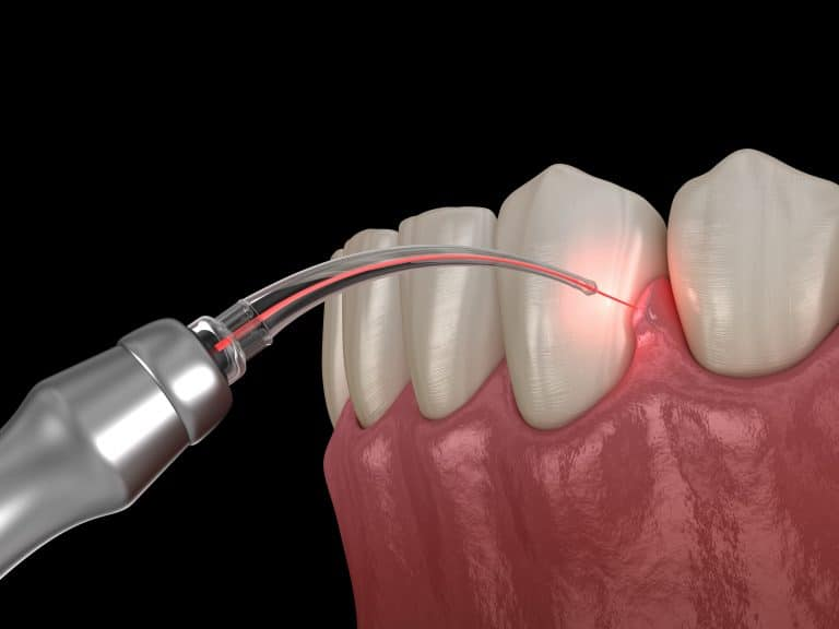
In dentistry, diode lasers are used only for soft tissue as they penetrate about 2 millimeters, perfect for the sensitivity of the teeth and mouth areas. Since dental surgeries often involve the gums, diode lasers are one of the most common ones used in dentistry. An image of diode lasers used in dental surgery is shown below. As discussed before, they cause less pain, better healing, and can lead to better technologies within surgical equipment.

A Diode Laser Cutting into Gum Tissue. Photo courtesy of Syosset Dentistry.
One of the most common uses for diode lasers is in crown troughing. Troughing is when gingival cells are vaporized in order to prepare for a prosthetic. Gingival cells are more commonly known as the gums. Traditional methods may take up to 5 minutes, while the laser can do it in under 60 seconds. The trough is not only more exact with a laser, but also cleaner. The heat from the laser kills the bacteria. This makes using diode lasers quicker, safer, and more hygienic.
Oral Surgeries
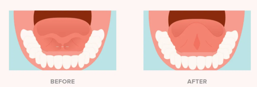
A frenectomy is any surgery that involves removing overlaying tissue. These are most often oral surgeries used for a tongue tie or lip tie. The image below shows a tongue tie pre and post frenectomy. These conditions are often not life threatening, and so dentists are hesitant to perform these soft tissue surgeries. This is due to the current surgical methods, since they have little hemorrhage control and there can be postoperative complications. Diode lasers can help make these surgeries more common, low risk, have cause little hemorrhage chances, and operational complications. No stitches are needed with lasers either, again making oral surgeries quicker, less invasive, and more exact. Diode lasers can even be used for procedures such as cold sores and canker sores. These procedures can even prevent cold sores from appearing.

A Tied Tongue Pre and Post Oral Surgery (Frenectomy). Photo courtesy of healthline.
The Biology of Diode Lasers in Tissue
The theory behind how diode lasers cut explains how these tools can revolutionize the dental industry. When the laser first touches the tissue, it is not immediately cut. The tissue has to heat up in stages, and with each stage molecular properties change. Tissue temperatures up to 90 degrees Celsius lead to coagulation and protein denaturing. Hemorrhaging, as discussed earlier with some oral surgeries, is prevented when coagulation occurs. Protein denaturing stops bacterial cells from splitting, and therefore spreading. From 100 degrees Celsius and up, vaporization of the cells occurs, and the tissue is cut. The vaporization leads to a fine surgical line and also sterilizes the cut tissue. All three stages together lead to a fine surgical cut, hygienic operations, and a relatively fast procedure.
These concentrated light sources are not yet the norm within dental offices. This is due to little knowledge on the benefits of lasers and few opportunities for dentists to learn. Hopefully this technology will grow within the next few years, and the ADA will start introducing diode lasers regularly into practices. With diode lasers becoming cheaper and easier to move, dental drills may become a surgical tool of the past.
