How often do we think about the importance of optical fiber probes in biomedical applications? When we think of optical engineering applications in the medical field, we often think of lasers, MRI machines, and imaging systems. And while these technologies helped advance medicine to a great degree, there are several other applications that present solutions to many of the challenges and barriers the field faces. One of these applications is optical fiber probes.
Optical fibers are widely known for revolutionizing communications around the world. They are the links that connect continents, countries, and homes to the internet. However, optical fibers have a huge impact on many other areas and fields, particularly the medical field.
In this article, we will discuss one very important application of optical fibers, an optical fiber probe developed for use in mid-infrared arthroscopy and joint disease diagnosis.
What is Osteoarthritis?
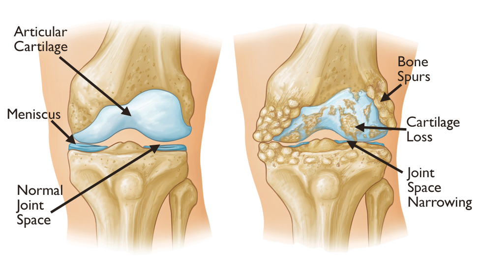
According to the CDC, Osteoarthritis also known as degenerative joint disease is the most common type of joint disease. It is a medical condition where the joints of the hands, knees, hips, and spine begin to deteriorate causing friction between the bones and sometimes the modification of the bone shape all of which manifest as pain or stiffness of the joints. Although this disease can affect any joint in the body, it is most common in the body parts mentioned above. A study conducted by Global Burden of Disease in 2019 found that nearly 528 million people around the world suffer from this disease. Osteoarthritis has become a source of major concern as these numbers are expected to rise as population aging continues to increase and other causes attributed to the disease such as obesity rates keep growing.

A side-by-side drawing of a healthy knee joint and a joint suffering from Osteoarthritis showing bone deformation and friction. Courtesy of OrthoInfo.
Diagnosis
In order to diagnose osteoarthritis in a patient and assess the status of the disease, arthroscopy is often performed. During this procedure, a thin tube attached to a fiber optic camera is inserted near the joint area through a small incision. The camera allows the surgeon to inspect the condition of the joint. However, despite it being a minimally invasive procedure, arthroscopy heavily relies on the judgment of surgeons who need extensive training to gain more skill and expertise. This can be especially critical for joints that are less commonly diagnosed such as the hip joints. Due to the risks/limits that traditional arthroscopy poses, researchers in the medical field have started joining efforts with engineers to design new medical equipment that can be used to assess osteoarthritis and other joint diseases in a qualitative way that ensures the reliability and accuracy of the diagnoses.
The Alternative
One of the proposed methods for diagnosing joint diseases is the Fourier Transform Infrared Spectroscopy or FTIR for short. The joint tissues that cover the bones and act as a liaison between different parts of the body have a very well-defined molecular composition. In fact, the concentration of the amides and the carbohydrates that form the tissue reflects its condition and in turn provides information about the development stages of the degenerative joint disease.
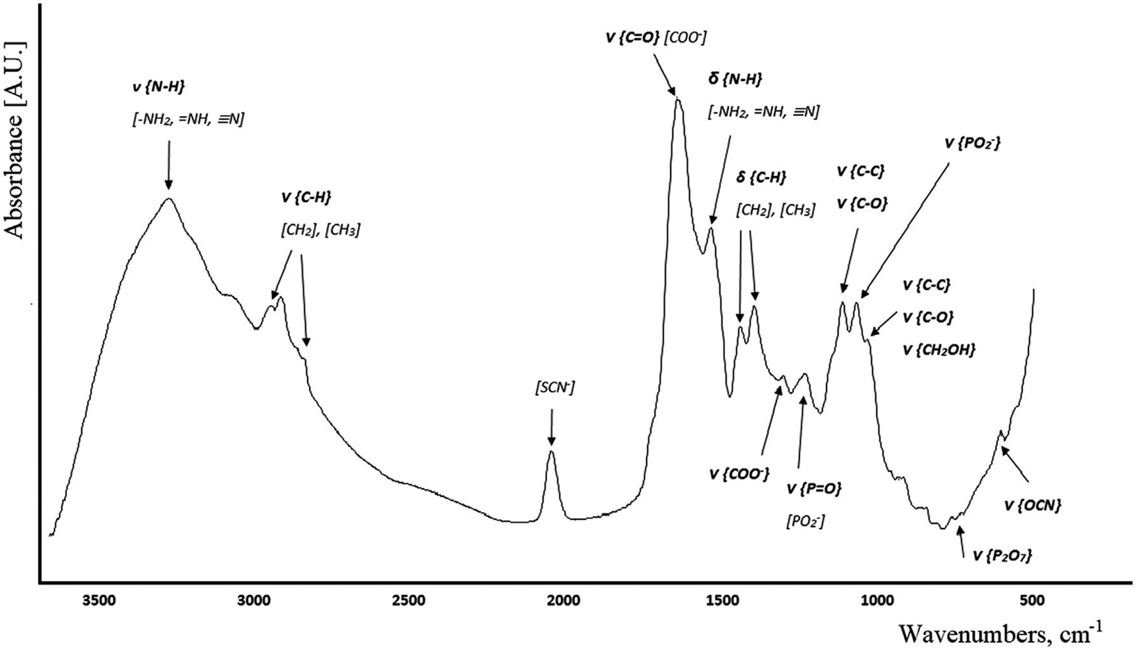
Mid-infrared light sources can be used to illuminate the tissue or the joint area. Each molecule has a unique frequency or region of frequencies known as the fingerprint vibrational frequencies. Depending on the wavelength, the light source excites vibrations in the different molecular structures that constitute the tissue and some of it gets absorbed. These excitations represent the composition of the material. Therefore, collecting the spectrum of light reflected by these particles will provide information on the composition of the joint tissue. As a result, the different levels of absorption will indicate the condition of the patient in a more objective and quantitative manner.

Example of an absorption spectrum collected via FTIR from the saliva of a healthy patient. Courtesy of the ScienceDirect.
However, with such a technique, the issue remains to find the adequate technology that would be able to illuminate the cartilage as well as collect the reflected light without causing damage to the surrounding tissue. The success and accuracy of this type of arthroscopy are reliant on the optical system used for measurement. Recently, engineers at art photonics GmbH have been working on designing optical fiber probes that are able to perform the aforementioned tasks all the while satisfying the rigorous clinical requirements at minimum noise.
Optical Fiber Probes
Concept
The optical fiber probes were designed to enclose 2 mid-infrared optical fibers. Optical fibers are excellent waveguides that can support a wide range of wavelengths from the visible spectrum down to the infrared with minimum propagation loss. Since the source wavelengths must be in the mid-infrared range, the optical fibers integrated into the probe must be special mid-infrared optical fibers. One of the 2 fibers is connected to a tunable infrared source and delivers the light from the source to the joint tissue where the sample will be irradiated while the other fiber collects the light reflected and feeds it into a unit that analyzes the data obtained, graphs it, and display it to the user.
Design Considerations
In designing the optical fiber probes to be used for MID-IR arthroscopy, the design team at art photonics GmbH had to consider various factors that will be discussed below. These include size restrictions, type of materials, and shape.
Dimensions
To meet clinical requirements, these probes were designed to be only a few millimeters in diameter, large enough to only support the mid-infrared optical fibers. Despite the size restrictions imposed on the probe, the fibers themselves cannot be thinner than 1 millimeter in diameter. The probe design uses polycrystalline fibers of a largest possible fiber diameter (1mm) so that the transmission of the system is enough to receive and emit light while maintaining a high signal-to-noise ratio.
Materials
The materials used to manufacture the probe have been carefully selected. It was required that these probes were made of biocompatible materials that are nontoxic and safe for use on human subjects. Additionally, it was a requirement that these materials could withstand sterilization by steaming. These requirements did not apply to the probe material alone, but to the optical fibers as well as the other components used, such as any integrated mirrors or type of reflective coatings. Polycrystalline Silver Halide optical fibers and Hollow Waveguide are known to be the best optical fibers for propagating infrared light in a >10um spectral range. According to studies, these materials are nontoxic and safe to use for in vivo measurements. However, by comparing the transmission spectra for the range of wavelengths needed for the FTIR spectroscopy procedures, it was found that the transmission of hollow waveguides is sensitive to bending. Given the high numerical aperture of Polycrystalline Silver Halide optical fibers and lower bending losses compared to hollow waveguides, Polycrystalline Silver Halide fibers were selected to carry the light from the source to the tissue.
Probe Tip
The probe tip is one of the most crucial parts of the entire assembly. Light collection/spectroscopy happens at the level of the probe tip. Therefore the geometry of the tip, as well as the material it is made of, are of extreme importance to the efficiency of the tool. Research has shown that using a diamond tip or crystal as the sensing element is very advantageous.
Diamond has a broad spectral response that includes the UV region, the visible region, and the far infrared. This makes it suitable for mid-infrared spectroscopy. Moreover, a diamond crystal has a large index of refraction which means that it does not have to penetrate deep into the tissue to collect data. By shaping the diamond tip into a prism, the penetration depth (or optical path length) is around 2 microns which is within the acceptable range for FTIR applications.
The properties of a diamond tip make it especially appealing for non-invasive procedures such as arthroscopy. Another reason that makes the diamond tip an excellent choice for this design is the fact that it can be chemically treated without deterioration. This allows for the reuse of the probe on many patients without the need to replace it after every procedure, thus further reducing the cost as well as waste.
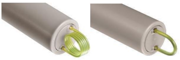
Fiber loop tips were also suggested and implemented in 5 different prototypes. In this configuration, only one piece of continuous fiber is used and at the end of the probe, the fiber is permanently looped or curved into a U-shape. This type of tip takes advantage of the properties of evanescent waves. These waves are known for being tightly confined in a straight optical fiber whose bending radius is infinite. However, as a fiber is bent, the wave starts leaking into the cladding and even coupling out of the fiber. The distance by which the evanescent wave gets displaced is known as the penetration depth and it is inversely proportional to the bending radius of the fiber. In other words, the smaller the bending radius, the larger the penetration depth and the farther light reaches into the tissue.

A picture of fiber loop probe tips. On the left, the probe tip is made of wound optical fiber. On the right is a U-shaped tip. Courtesy of art photonics GmbH.
Unlike diamond tips, fiber loop tips are cheap, and they can reduce the cost of production substantially.
Shape
When it comes to the shape of the probe, the engineers at art photonics GmbH proposed 2 designs. The first one consists of a straight shaft with a tip attached to its end. On the other hand, the second design had a straight probe with a tip forming a right angle (90-degree angle) with the shaft. This design was referred to as the hook-shaped probe. Several prototypes having different tips were developed and compared. In the section below, the results of the performance comparison tests will be presented.
Probe Testing
In order to test their prototypes, art photonics GmbH collected the transmission spectra for the different designs and evaluated their performance. Based on the transmission rates obtained, it was clear that the diamond probe tips, although more costly, perform better than fiber loop tips. As explained in a previous section, fiber loops are obtained by bending optical fibers which inherently suffer from bending losses. For very small bending radii, these losses cannot be ignored. It is worth mentioning however that the signal from the fiber loops is stable and generally, fiber loops satisfy the clinical requirements. IR Medtek is a good example of a company developing a diagnostic technique using the fiber loops. IR Medtek LLC, a start-up commercializing a fast, non-invasive, and accurate cancer detection probe, utilizes mid-infrared spectroscopy to determine molecular changes unique to cancerous tissue. This technology was developed at The Ohio State University and the James Cancer Hospital, Columbus, OH USA. IR Medtek’s first target market is a skin cancer detection system which uses fiber loop probes provided by art photonics, GmbH. The goal is to provide the medical community with information to enhance treatment decisions and eliminate the bulk of negative biopsies which are intrusive to patients and taxing on the medical care system.
Finally, given the very low cost of the fiber loops, they are designed for single use only and are easily detachable.
Results
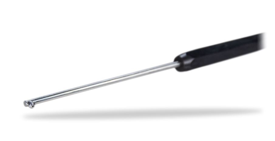
The various tests and designs that art photonics GmbH has developed led to the optimization of the diamond tip probe, which is compact, robust, safe for medical use, and has a high transmission over the required IR range. It is also worth mentioning that this technology allows for the evaluation of a tissue in real-time with a recording duration of about 1 minute per spot.

A picture of the hook-shaped diamond tipped optical fiber probe. The shaft material is stainless steel. Courtesy of Art Photonics/Miracle.
Conclusion
Efforts of developing optical fiber probes to improve arthroscopy techniques could allow for the early diagnosis of Osteoarthritis. Early diagnosis is very critical for various reasons. It has the potential of reducing the financial strain on the healthcare system, which in the United States alone would reduce the costs by $16 billion per year. Identifying the early signs of the disease which unfortunately cannot be performed using ordinary imaging techniques would also allow early access to treatment and lower the risk of disabilities or impairment, especially among older adults. Additionally, the evolution of this technology can increase the diagnosis accuracy making it more reliable.
If you liked this article, you might also be interested in our latest blog post where we discuss the role of fiber probes in process analytics.
