Currently there is development in the biomedical sphere surrounding a very well documented species: Zebrafish. One benefit of studying these fish is the insight on neural development literally visible in their transparent larvae stage. However, these are small fish; with eggs 0.7 mm in diameter and larvae about 3 mm in length. As expected, researchers use powerful and accurate visual tools to properly study this larvae’s development. Multiphoton microscopy on zebrafish allows for proper imaging and study of the live transparent fish larvae. We will discuss this process today in this article.

Example of multiphoton microscopy on zebrafish embryo. Image courtesy of poyltechnique-insights.com
What is Multiphoton Microscopy?
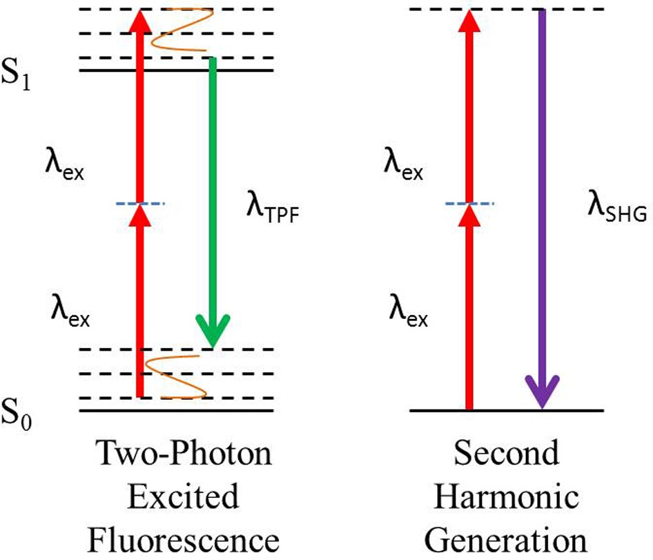
Fortunately, we have a few other articles on this blog which explain the process much more in depth. Two articles in particular can better educate on the topics of two photon microscopy applications and fluorescence microscopy techniques. However to summarize: multiphoton microscopy involves emitting a photon dense laser at a light sensitive substance. This results in the substance reacting fluorescently. The substance then emits a photon in return with a different wavelength from the photons from the laser.

Demonstrating 2 kinds of multiphoton microscopy. One with decay depending on material and another with a return wavelength exactly double the wavelength of incoming photons. Image courtesy of Nature.com

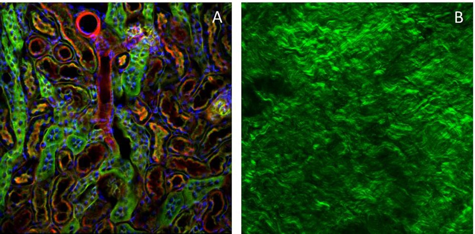
Demonstrating the results of both methods of multiphoton microscopy. Note the variation in color depending on substance in A contrasting B’s monochrome coloration. Image courtesy of Nature.com
As shown in the images, multiphoton microscopy is a method which returns a colored image from stimulated fluorescence. The resulting image can achieve cellular resolution easily while non-invasive in the sense that no incision is required. This technique is incredibly useful for biomedical applications due to its non-invasive nature and impressive depth capacity.
Multiphoton microscopy on zebrafish allows for detailed study and accurate observation in experimental settings. These observations have produced so many discoveries and developments in the field of medicine and biology.

Results of 2 photon imaging of zebrafish neural population activity. Image courtesy of ScienceDirect.com
Why do we study Zebrafish?
Zebrafish are both convenient to study and provide a biomedical research “model species” for a number of reasons as follows:
- Zebrafish share about 70% of human genes
- It is convenient and well known how to breed and house zebrafish (requiring little space)
- External eggs and fertilization provide ease in manipulation/documentation of embryos
- Transparent larvae allow researchers to study developing neural structures of living organisms
These reasons and more are further discussed in detail at the following microscopemaster.com article

Various changing florescence allow for imaging of completely different organ groups at cellular resolution. Image courtesy of ResearchGate.com
Demonstrated above is selective imaging of interest organ groups possible in zebrafish. Multiphoton microscopy on zebrafish permits extremely good imaging and documentation of neural network structures in particular.
What are we studying?
While the list of studies surrounding zebrafish is rather extensive, we will be focusing on discussing studies that utilize multiphoton microscopy on zebrafish.
One study, outlined in this video from JoVE, introduces a light responsive agent at an early stage post egg fertilization. This produces a live genetic landmark which can be viewed through 2-photon microscopy at a cellular resolution. Following this, the larvae are then grown in a low melting point agarose to keep the embryo immobilized. Finally, the zebrafish larvae can then be properly observed using 2-photon microscopy at a desired focal length/depth. The observation process, once started, can be done at any point in the development of the larvae.
In addition, the process can generate multiple layers of recorded data by creating plane images at different depths. Repeated observation produces what is known as cell fate mapping. Cell fate mapping is when researchers follow a targeted and marked cell/collection of cells to determine what it becomes over time. Cellular marking passes on to the cells’ descendants in order to further determine what each becomes after development.

Zebrafish embryo is held still while multiphoton microscope begins scans to produce high resolution images of a live specimen. Image courtesy of JoVe.com
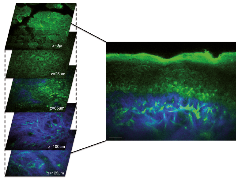
Performing multiphoton microscopy on zebrafish allowed researchers to develop recordings of brain functions at cellular resolution using 3D scanning. A process of scanning multiple planes with a multiphoton microscope created the 3D image. Each plane has a focus distance a small increment away from the previous plane. Sufficiently iterating this process produces a high resolution 3D scan. Refer to the image below which exemplifies the technique (also noting that the planes are only about 25 micrometers apart).

MPM example of layering to create 3D image made from human skin scans. Image courtesy of Medicaljournals.com
Results of Multiphoton Microscopy on Zebrafish
The results of using multiphoton microscopy on zebrafish have given us a deeper understanding of living organism’s development and neural structures. Working with the zebrafish has given researchers many flexibilities in biomedical applications. Notably the contribution to neural structure development and disease treatment have been immeasurable in developing medicine. One drawback to working with zebrafish over mammals like mice is a lack of mammalian organs (e.g. Lungs, mammary glands, hair, etc.). Medical researchers have utilized zebrafish to model diseases of interest with the intention of developing treatment to scale up for human use.
Properly modeled diseases in zebrafish include: Duchenne muscular dystrophy, human melanoma, and human neurodegenerative diseases.
Conclusion
To conclude, multiphoton microscopy on zebrafish remains a developing methodology employed by researchers today. The benefits of these non-invasive and deep scanning images have already assisted in the modeling of diseases and development of adequate treatments. Human understanding of biological and neurological development increases because of research conducted on zebrafish. Use of multiphoton microscopy on zebrafish is a novel methodology that furthers this understanding with unprecedented 3D scans. For this and many other reasons advancements in this realm of scientific research have many investments and observers.
This blog post is brought to you by RPMC Lasers Inc, a leading supplier of laser technologies in North America.
Further Readings
Related articles on multiphoton microscopy:
https://www.findlight.net/blog/2020/02/23/fluorescence-microscopy-multiphoton-and-other-techniques/
https://www.findlight.net/blog/2020/06/21/two-photon-microscopy-and-diagnostic-applications/
Uses of zebrafish as biomedical model research paper
Article of zebrafish in research + general info
Multiphoton microscopy explanation article
Full brain scan using MPM on Larvae research paper
Science direct 2-photon imaging of zebrafish
