In modern medicine, the ultrasound is a go-to method for viewing the body’s internal structures, given its non-invasive nature. Ultrasound produces images in real time by relaying high frequency soundwaves through a special probe. The soundwaves echo off different organs and tissues and return to the probe. A 2D or 3D pixel then forms based on the echoes respective distance. This is called “pulse-echo ultrasound” since it pulses waves in quick succession and creates a fully detailed image from the echoes. Optical ultrasound produces these soundwaves differently, but has some key advantages.
All-Optical ultrasound, instead uses a pulsed light to emit soundwaves via the photoacoustic effect. The photoacoustic effect is when light produces heat, resulting in increased pressure that is expressed in the form of a soundwave. From there, a transducer measures the returning soundwave in the same way as traditional ultrasound. It may seem redundant to use light to recreate pulse-echo ultrasound. However, traditional ultrasound requires a probe containing a piezoelectric crystal, which gives off soundwaves and receives the echoes for translation. While they are useful, probes with a piezoelectric crystal emit electromagnetic interference that prevents them from being used near radiology devices like MRI or CT. It also limits their use in electrophysiology, neurological treatments and other treatments involving electricity. In many procedures involving radiology, live ultrasound imaging could be of great benefit.

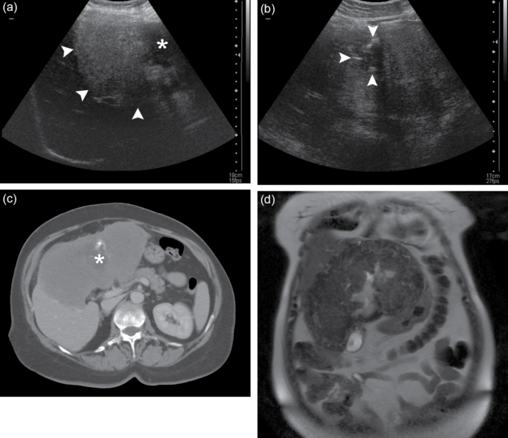
Pictured above is a comparison of a ultrasound and CT images in a guided needle biopsy. This procedure is to identify an abnormal growth, in this case a parasite, that has been growing in a patient’s abdomen. It serves as a good example that multiple images provide the best diagnosis. The arrows in the ultrasound reading to the top point to a solid mass growth. The CT scan on the bottom is then used to give further detail. Photo retrieved from Wikipedia.
How Optical Ultrasound Works:
A recent work from University College London presents the first all-optical ultrasound to produce an image in real time, with comparable video quality and signal-to-noise ratio. Other optical ultrasound configurations exist but require much longer scan times that limit their use in the laboratory. An optical setup uses a galvo mirror instead of the piezoelectric crystal, eliminating the electrical component. With a galvo mirror, light reflects to many different points along a tissue structure to capture movement in real time. The resolution is only limited by the inertia of the galvo mirror, its ability to handle a rapidly pulsed laser, and the aperture size.
Electronic probes in traditional ultrasound analyze a specific resonance. For this reason, probes come in different forms suited to specific applications. There are some multi-purpose probes that handle a range of tasks, but intense studies like cardiology require specially made probes. These issues require waves that can reach structures located deep within the human body, like specific heart arteries or malformations on pelvic structures for women’s health. Similarly, the wavelengths in a single optical ultrasound probe are tunable to adjust scan depth and resolution for various purposes. Instead of switching between multiple probes, an optical ultrasound could simply be adjusted to different settings for all-in-one capability. The benefits do not stop there, as optical ultrasound provides greater variability than the standard ultrasound.
This configuration opens the possibility of using bundled waveguides to make a longer and more flexible device for ultrasound imaging. As previously mentioned, the optical probe and transducer can be used separately from the electrical parts. This opens ultrasound’s use case in conjunction with electromagnetically intense tools like MRIs and CTs. The ways in which these can be used together to impact care are discussed below.
Applications in Care:

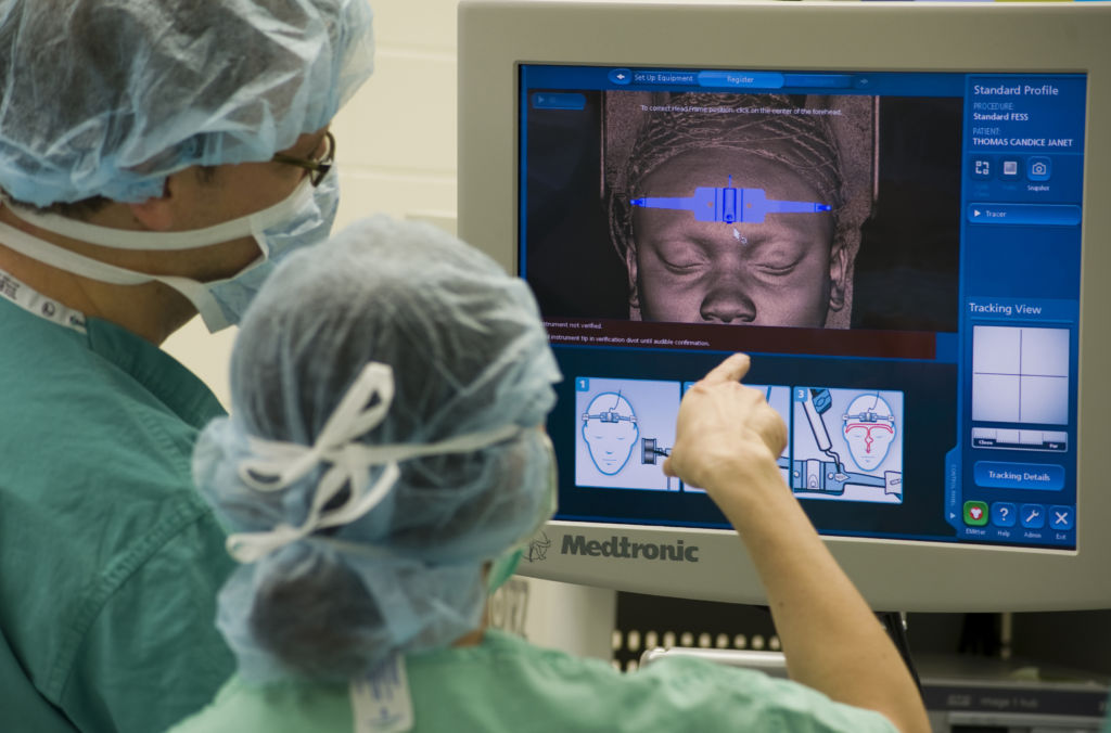
Pictured above are two surgeons analyzing a CT scan to assist in their treatment. The CT machine and monitor emit electromagnetic interference, which throws off the reading of an ultrasound. Fully optical ultrasound uses a probe with no electrical components, so that both CT and ultrasound can be used in conjunction. Photo courtesy of Bobby Jones from Joint Base Andrews.
Vascular access procedures involve inserting a catheter to re-open a clogged coronary artery. It requires accurate guidance, with help from both CT and ultrasound. Typically, ultrasound identifies veins that are large enough for catheter insertion during preparation. During the procedure, CT scans or an x-ray guided catheter provides live imaging as it navigates the body. Cardiac CT machines are extremely expensive, and use twice the amount of radiation as a normal CT. There are some methods in which surgeons have experimented using only ultrasound in catheter insertion. These are not developed methods, and the procedure would be more safe and more effective if ultrasound could be used with a plurality of imaging techniques. In addition to being more costly and potentially damaging, MRIs and CT scans are highly invasive. Minimizing the need for these scans with optical ultrasound can certainly increase the patients’ comfort and safety.
Diagnostic cardiology could greatly benefit from this technique as well. Overall, it is expensive and not feasible to consistently test a patient’s heart for potential vulnerabilities because of the factors listed above. Detailed optical ultrasound would be a good supplement to CT angiography to get a more detailed picture without resorting to cardiac CT.
Another area where CT scans have been proliferated is to study the abdomen and pelvis. These include women’s health (OBGYN), urology and various forms of oncology. The most obvious of these is ultrasound in obstetrics (OB), or the study of childbirth. Particularly, ultrasound is used to produce an image of the child while still inside the mother and identify any irregularities. However, imaging the pelvis is extremely important to both men’s and women’s health, especially for surgeries. Examinations on the stomach, intestines, kidneys, liver and prostate use both CT’s and ultrasound. In some cases, they require endoscopy. Searching these areas for abnormalities that indicate cancer require real time detailed images, along with a physician’s trained eye. Once again, optical ultrasound would provide video rate observation to identify growths in these areas with non-invasive methods.
Conclusion:
The specialties that could benefit most from this innovation include pulmonary medicine, cardiology, OBGYN, urology, pelvic medicine and emergency medicine. These are all areas in which both optical and radiological tools are used for treatment and diagnosis. Coincidentally, they all involve deep, difficult to reach structures in the body which entail invasive procedures to get a good look. Ultrasound without electromagnetic emissions means no hassle for specialists who use CT and MRI imaging in conjunction with ultrasound. This lets surgeons in the operating room bridge live motion capture ultrasound, with immediate scans. Surgeons can easily guide instruments like a catheter with the help of ultrasound. In the case of endoscopy, this technology may even do away with the need for the insertion of a camera to get live imaging. While radiology is still required to reach positive outcomes, optical ultrasound can help reach a diagnosis quicker, with less harmful methods.
References
- Erwin J. Alles, Sacha Noimark, Efthymios Maneas, Edward Z. Zhang, Ivan P. Parkin, Paul C. Beard, and Adrien E. Desjardins, “Video-rate all-optical ultrasound imaging,” Biomed. Opt. Express 9, 3481-3494 (2018)
- Brian W. Pogue, “Optics of Medical Imaging” SPIE Newsroom, 22 January 2018. Accessed 9/9/2018, via http://spie.org/newsroom/optics-of-medical-imaging?SSO=1.
