Search Results for: medical imaging
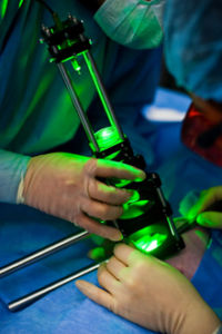
Optical Fibers for Medical Sensors and Light Delivery
Optical fibers are a convenient instrument for robust light transfer. Their low losses have enabled their wide use in long distance telecommunication systems while service providers like AT&T, Verizon ...
Computational Ghost Imaging: An Unconventional Solution
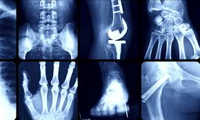
X-Rays and Imaging We commonly associate x-ray imaging with hospitals and broken bones, but why are x-rays the chosen source of radiation instead of visible light or electron beams? The high initial energy ...
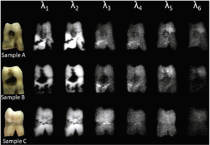
Dental Caries Diagnosis Using Fluorescence Imaging Techniques
A research team from the Military Technical College and from Cairo University in Cairo, Egypt have developed a fluorescence hyper-spectral imaging system which can be employed for diagnosis and detection ...
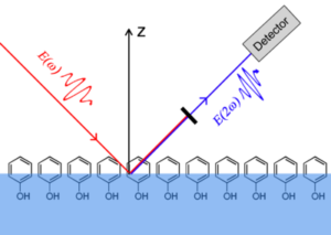
Nonlinear Imaging to Characterize the Microenvironment of Tumors
Developing a clear characterization of a tumor’s microenvironment is imperative in understanding its progress. A new approach to determining cancer’s development involves a thorough study of ...
Medical Lasers: A Guide to Various Effects and Choices
The first medical application of lasers was reported by Goldman in 1962. In cardiovascular surgery, McGuff first used a Ruby laser in 1963 for the experimental ablation of atherosclerotic plaques. After a ...
Neural Imaging with Visible Light: Advanced Implantable Sensor
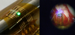
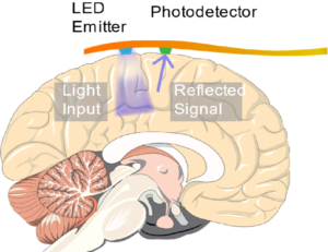
Previously, we discussed a technique for producing high resolution maps of neural activity in the brain called Optical Recording of Intrinsic Signal, or ORIS. Conventional methods for performing ORIS ...
Neural Imaging with Visible Light: Implantable Optical Sensors
Previously, we discussed how visible light in combination with implantable optical sensors was used to map epileptic neural activity and localize focal seizures on the surface of the brain. Scientists at ...
Machine Learning Enables OCT Imaging of Optic Nerve Head
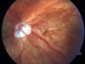
Medical imaging of the optic nerve head (ONH) is immensely important to study vision disorders like nearsightedness and glaucoma. Doctors traditionally use optical coherence tomography (OCT scans) to ...
Calvarial Fracture Imaging Using Raman Spectrometry
Scientists from the City University of Hong Kong’s Department of Physics revolutionized calvarial fracture imaging through the use of Raman spectroscopy. By allowing these defects to be imaged, the ...
Endothelial Surface Layer Imaging: Diagnosing Early Sepsis
A joint team of researchers from the Korea Advanced Institute of Science and Technology and the Seoul National University have achieved in vivo imaging of the endothelial surface layer. The endothelial ...


