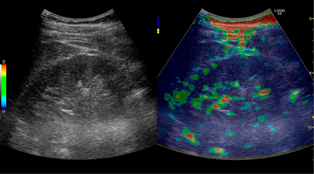
Elastography allows for many insights into the properties of tissues and tumors. It can be achieved through various imaging techniques, such as ultrasound, pictured above. However, in recent years, research has been done into the potential of photoacoustic imaging as an alternative to the use of MRIs or ultrasounds for such images. Image courtesy of Wikimedia Commons.
Introduction to Elastography
Elastography is a type of imaging modality that provides information on the elastic properties and stiffness of the tissue being reviewed based on spatial distribution. It is most commonly used when imaging livers to test for fibrosis or steatosis- fatty liver disease. Fibrosis leads to a buildup up scar tissues, and therefore, researchers look for signs of abnormal stiffness to make a diagnosis. Elastography is also used in tumor diagnosis, as cancerous tumors are often harder than the surrounding healthy tissue. Finally, in some less common cases, elastography is used for musculoskeletal imaging, to asses the state of muscles and tendons.
Ultrasound imaging and Magnetic Resonance Imaging (MRI) are most commonly used for elastography. Optical coherence tomography (OCT) and computed tomography (CT) imaging have also been explored as potential techniques for elastography. In the last decade, photoacoustic imaging (PA) has also been identified as another possible imaging technique. These imaging techniques allow for diagnosis without the use of biopsies or manual palpitation. Biopsies are largely invasive, and manual palpitations relies heavily on the medical practitioner’s knowledge and experience. Additionally, with manual palpitations, the patients runs the risk of small or deep tumors being missed. Therefore, for many types of tumors, elastography, regardless of which imaging technique is used, is much preferred to the older diagnostic tools of biopsies and palpitations.
Introduction to Photoacoustic Imaging
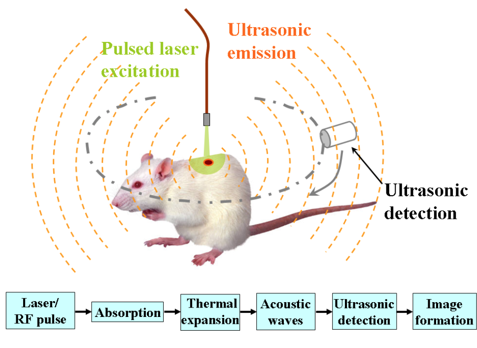
Photoacoustic imaging is a form of imaging that employs both photonics (light) and acoustics (sound). During imaging, short non-ionizing laser pulses excite acoustic pulses. This occurs as some energy provided by the lasers will be absorbed. Absorbed light is then converted to heat, leading to a thermal expansion and emission of wideband ultrasonic waves. This is shown in the simple schematic below.

This simple schematic details how the use of a laser source can lead to acoustic wave detection. This process allows photoacoustic imaging to work successfully. Image courtesy of Wikimedia commons.
Measuring acoustic waves instead of light waves is desirable as acoustic waves scatter and absorb less, and therefore provide better insight at high depths within the tissue. Both 2D and 3D images can be made using photoacoustic imaging.
The most widely used for of photoacoustic imaging is photoacoustic microscopy (PAM). Imaging depth is modified in experiments by optimizing lateral resolution. Resolution changes with changing optical and acoustic foci.
Photoacoustic Elastography Experimentation
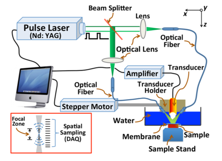
The first elastography images made using photoacoustic imaging were studied in 2011. Since then, initial progress utilizing photoacoustic imaging was slow but has started to pick up in the last few years. In one experiment reviewed, an acoustic-resolution (AR) PAM system was utilized. In such a system, high lateral resolution can be achieved through increasing the center frequency of there ultrasound transducer and through tightening the acoustic focusing. AR PAM systems general have a lateral resolution of 15 to 50μm. The schematic of this system is shown below. Important components of this system include the short-pulse lasers, ultrasonic transducer, and optical fibers.

Shown above is the schematic diagram of a photoacoustic microscopy imaging system, which was used to produce photoacoustic elastography images. Diagram courtesy of SPIE Digital Library, Journal of Biomedical Optics.
Experiments were performed on agar base tissue-mimicking phantoms first, as the physical characteristic of these phantoms were manufactured by the researchers themselves. After software was optimized to best interpret and display elastography results, AR PAM was used to image a mouse leg before and after applied compression. Ultrasound was also used so as to compare the results between the two imaging modalities.
Results & Conclusion
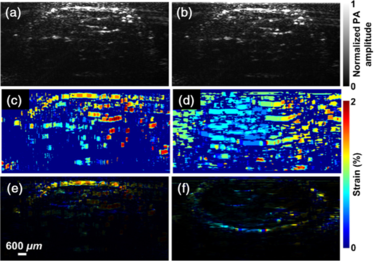
The resulting images obtained through mouse experimentation are shown below. Researchers identified several challenges associated with photoacoustic elastography imaging. First, there existed low photoacoustic single contrast. This was accompanied by a difficulty regarding discrimination of contrast in the signal due to variations between elastic property and other parameters. Finally, quantitative measurement of the elastic properties of tissue was a challenge.

The figure above display the results of imaging a mouse leg before and after applied compression. Image (a) shows a photoacoustic elastography image of the leg before compression while (B) shows the leg after compression. Images (c) and (d) are strain images of the in vivo mouse leg obtained by PAE and ultrasound elastography (USE) respectively. Finally (e) displays strain image obtained by PAE superimposed on the structural PA image while (f) displays strain image obtained by USE superimposed on the structural ultrasound image. Image courtesy of Spie Digital Library, Journal of Biomedical Optics.
These challenges can be overcome with more research and development. External mechanical stimulation of tissues was already proposed by researchers to overcome low signal contrast. Additionally, existing technologies used in ultrasound imaging for quantitative recovery of elastic properties could be modified for PAE.
Despite these challenges, PAE is still a very viable form of elastography, especially considering its youth. Rapid progress could occur in the next decade to make it competitive with MRI and ultrasound based elastography techniques. Ultimately, it will allow for improved tissue and tumor diagnosis as well as improved patient care.
To read more about discoveries being made regarding photoacoustic imaging for the purpose of elastography, an article can be found here.
