Multi-photon microscopy is a useful tool for analyzing in-vivo interactions between photons and living tissues. One of its characteristics is deep tissue penetration, meaning it can get a better look at vital organs or structures within the body. Multi-photon microscopy relies on two photon excitation of the studied samples with a long wavelength laser. Longer wavelengths indicate lower photon energy causing limited to no damage to the living tissue. However, photon microscopy has a limitation that hinders its use in neurology where there are high concentrations of neuronal calcium. A uniform distribution of photons illuminating the sample is spread over a quickly growing area. When this happens, several noise sources interfere with the sensor.
This leads to two outcomes. First, the brain’s high concentration of calcium makes finding photons harder for the microscope, leading to a poor image. Second, the process produces a massive data stream that can be difficult to extract a live image from. This means long imaging times, and also a delay in measurement of extremely fast neurological responses. Researchers isolate each photon detection event to limit fluctuations in brightness from a data skew. The solution lies in time stamping each photon detection event to sequentially put a threshold on each event’s tolerance for light disturbance.

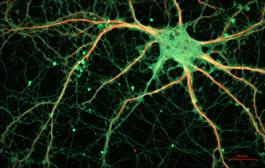
Pictured above is a neuron in the hippocampus of a rat. Photon counting is used to measure specific electrical responses sent through these neurons. Picture courtesy of Zeiss Microscopy, on Flickr.
As previously mentioned, wide expanses of photon deprived neuronal calcium in the brain require a much faster microscopy process than is achievable by most multiphoton microscopes. Photon counting is a complex experimental method to improve multi-photon microscopes in deprived conditions. This method uses a pulsed laser to determine a photon’s brightness and arrival time. Photon counting is effective for imaging in the brain, but it requires electronics experience, expensive parts and plenty of time. A new software design hopes to make photon counting quicker and easier to perform.
How does the software make photon counting more effective?
New research out of Tel Aviv has produced an open source plugin for existing microscopes. It is specially designed to handle microscopy in photon deprived conditions, like those in neurology. It includes a data efficient way to implement time stamped photon detection. The idea behind their creation is to easily allow in-vivo imaging of the brain and its neurological responses on just about any multi-photon microscopy imaging system. All this without requiring complicated electronic configuration by the users.
The limit to other methods of photon counting in neurology is that the data collection time scales with the area of samples taken, rather than with the number of detected photons. This means that these systems are always collecting data on brightness, even as they measure areas with none. This prolongs the scan time in addition to disrupting the image with a skewed data set. Essentially, the signal detected from a signal photon is buried in the noise and the resulting image has trouble differentiating between a single photon detection and the noise. Similarly, the rare occurrence that multiple photons are detected in a single cell throws off readings with its high peak brightness.
This photon counting software collects only the exact time that the photon event occurs. This way, the software does not have a massive inflow of photon-less data when it passes over neuronal calcium in the brain. Of course, time stamping photon detection is not the only problem to solve. This only places the events in a discrete list for easy reference from the software. From there, the arrival time of the photon can be measured without needing to reference volume or size of the measured area. Other photon counting software has been unable to timestamp photon detection in real time. Delays and insufficient memory plagues these methods, making neurology a challenge for standard photon counting.
How does it compare to other photon counting methods?
The program nullifies the effects of empty cells and optimizes detection time since it doesn’t process unnecessary data. This technology allows for a host of benefits. The first of which is faster imaging and the capability of lasting much longer. In addition, the method doesn’t suffer from any resolution loss no matter how long the scan time lasts. With an improved data stream, the researchers were able to test on the neuronal tissue of fruit flies. They imaged a 3D rendering of a fruit fly’s response to odor with almost 200 times less data streamed than other systems measuring a similar area. Measuring these responses in real time gives scientists a new way to study the brain’s reaction to trauma or various diseases/disorders.
Essentially, the device optimizes photon microscopy so that no two scanning elements are overlapping in their use. It limits overlap by time stamping each photon event independent of total volume scanned. After the scan, the software find’s the photon’s brightness and subsequent arrival time (and thus the origin). From there, the software can generate a 3D picture based on these arrival times. Continuous 3D imaging like this is big for in-vivo diagnostics, especially when done during a procedure. Other methods of photon microscopy haven’t adapted to the various photonic conditions found in organs like the brain. Using area-based detection leads to over-utilizing the scanners in a traditional photon microscopy.
Conclusion
Photon microscopy has shown great benefit to neuroscience in a number of ways. Imaging on a photon scale gives insight to quick bursts in a synapse and long-term memory. The new device makes it easier to analyze larger volumes of brain matter with less data usage. This opens up larger study capability to more labs, but also allows people to capture quick events like a neurological response in three or more dimensions. The speed of an olfactory response in a fly makes any sort of delay in imaging time ruinous to results. The technology marks an exciting accomplishment in computer science with microscopy, as it is an application engineered for use by people without electronics experience.
Despite its benefits, photon microscopy is still a field with high barriers to entry because of expensive technology. Not to mention, the field requires an intersection of knowledge across life sciences, optics and software to get a look at complex in-vivo cases like the brain. One of the technology’s most striking features is that it is an open source software package. This makes it easily to install without prior technical knowledge. Simplicity is great to help labs perform the more complex diagnostic tests without needing the help of an experienced consultant. Neurological tests of all kinds are widely available, and easier to perform in more labs.
