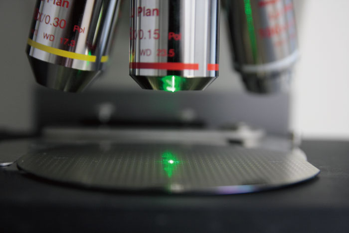
Raman microscopy utilizes light scattering to observe molecular vibrations in a sample. Courtesy of Gaia Science.
Introduction to Raman Microscopy
Raman microscopy is a laser-based spectroscopic technique used to observe molecular vibrations. It is frequently utilized in chemistry to provide a “fingerprint” of different molecules and insight on the composition of a sample. Raman microscopy gets its name from the Indian physicist C. V. Raman, who observed inelastic light scattering properties in liquids during the 1920s. Raman and his student, K.S. Krishnan, successfully detected nearly sixty different liquids and gases using Raman spectroscopy. He later received the Nobel Prize in Physics in 1930.
However, it took several years before technological advancements allowed for effective and sensitive Raman-based instruments. While initial experiments resorted to photography for recording results, the technology was significantly improved with the invention of the laser in 1960. Utilizing a laser allowed for focus on very small sample sizes. It also improved the detection power of the microscope and allowed for detection of low frequency molecular vibrations. Today, diode lasers are frequently used in Raman instrumentation for increased reliability and accuracy in sample measurements. It is also commonly combined with other complementary analysis methods such as scanning probe, scanning electron, or confocal laser scanning microscopy, to provide a more thorough understanding of a sample.
Currently, the market profile for Raman microscopy includes pharmaceuticals, academia, semiconductor, polymer and biotechnology industries, and government applications. This growing market is driven by recent technological advancements in microscopy, such as high-throughput and high resolution techniques. Increased research in fields such as neuroscience, nanotechnology, and material sciences also drives the growing market.
Operating Principle
Raman microscopes utilize spectroscopic techniques coupled with a standard optical microscope to visualize a sample. This technology relies on a concept called Raman scattering, which describes the method in which photons scatter by molecules at higher energy levels. This concept is unique in the fact that when photons scatter in this manner, the energy of the particle is not conserved.

A blue laser used in Raman spectroscopy. Courtesy of University of Cambridge.
Excitation lasers in the visible, near infrared, or ultraviolet light range interact with molecular vibrations and rotations. This interaction results in the laser photons shifting up or down in energy. The energy shift provides information on the various vibrational modes in the system, which are specific to different chemical bonds and molecular symmetries. Properties of a material are determined through analyzing the energy shifts and/or comparing them to a known library of wavelength spectra to identify a substance.
A typical Raman microscope includes a standard optical microscope, an excitation laser, monochromator, and a sensitive detector. Several variations of Raman microscopy including confocal direct imaging and hyper-spectral imaging are available. Direct imaging is a method in which an entire field of view receives a small range of wavelengths from the excitation laser. This technique is beneficial for recording the distribution of a specific molecule throughout a sample. In comparison, hyper-spectral imaging collects and processes information from the entire electromagnetic spectrum. Thus, the distribution of several molecules may be recorded.
Overall, Raman microscopy offers high spatial resolution and the ability to image individual cells on the sub-cellular level. It is non-destructive and non-invasive, so measurements can be taken without damaging a sample. Additionally, water does not tend to interfere with Raman spectral analysis, so it is suit to analyze minerals, cells, proteins, and other biological materials.
Applications
Raman microscopy finds a wide array of applications in varying fields. It offers many different advantages that make the technology extremely versatile. Although most frequently used in chemistry, this technology also finds applications in art, geology, defense, biology, and many more.

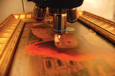
Raman microscopy can analyze the materials used in different artwork and assess authenticity. Courtesy of Art New England.
One example of the widely diverse applications of Raman microscopy are its benefits to the fine art and historical industries. Because it is non-destructive and non-invasive to samples, Raman microscopy offers an efficient method in analyzing historical documents, ancient works of art, etc. By identifying specific molecules, it can also provide insight to social and economic conditions at the time of a piece’s production, or help determine methods of preservation for such documents.
Furthermore, it is extremely helpful in determining forgery. Visualization of individual pigments provides information on degradation and the types of materials used in an artwork. Raman microscopy identifies the various types of materials present, which experts then cross-reference with the types of materials available during the artist’s lifetime. This can give clues to whether a piece of art is real or fake.
In geology, it is used for gemstone and mineral identification along with determining mineral distributions in rock sections. Geologists commonly require characterization techniques that can provide detailed information on rock formation history. Raman microscopy provides chemical identification, characterization of molecular structures, and effects of bonding, making it the perfect tool for geological studies.
Lastly, this technology is increasingly useful in homeland security. The ability to detect explosives accurately, quickly, and sensitively has become progressively important at airports, train stations and federal buildings. Moreover, Raman microscopy is currently in experimental stages for detecting residues involved in the construction of explosives.
The Future of Raman Microscopy
Raman microscopy provides an extremely sensitive and versatile tool that can identify various compounds based on their molecular vibrations. This technology is useful in many different fields including art, history, geology, defense, and more. The invention of the Raman microscope has opened up the door to endless research opportunities and has provided a tool for scientists, doctors, and professionals throughout the world. There is a growing interest for efficient and sensitive imaging equipment. This correlates to a projected forecast of a $1.8 billion market for Raman microscopy by 2021.
