Introduction
Raman spectroscopy stands as a cornerstone in the realm of vibrational spectroscopy boasting its unique capability to reveal the molecular intricacies of diverse materials and biological systems. By analyzing the light scattered from a sample, this technique provides information about molecular structures. This analytical tool has become instrumental in studying biological landscapes and evaluating nanostructured materials, forming the basis of fundamental advancements in understanding molecular compositions, interactions, and behaviors.
Raman spectroscopy enjoys a myriad of applications in various fields including material sciences, and even food and agriculture. However, with the convergence of technological advancements and its capability to provide detailed information on molecular composition, Raman spectroscopy was rapidly integrated into the medical and biological sciences. Its non-invasive nature and high accuracy in analyzing molecular signatures made it an invaluable tool in early cancer detection. In this article, we will explore the operating fundamentals of Raman spectroscopy and its importance in the medical field.
History
Raman spectroscopy was named after the Indian scientist Sir C.V. Raman who discovered Raman scattering. In 1938, the researcher observed that when polychromatic light traveled through a liquid, a fraction of it was scattered. He studied what became known as the “Raman effect”. This is a phenomenon that depends on the nature of the particles constituting the material. On the microscopic level, light incident on a material will interact with the bonds of molecules forming that material. Upon interacting with the incident light, the vibrating molecule re-emit the light in an arbitrary direction. A portion of this scattered light undergoes a shift of energy or frequency as a result of that interaction which corresponds to the unique bond of that molecule. As such, the Raman effect allows the identification of elements in molecules.
Soon after that, scientists started exploring applications that take advantage of the Raman effects, and so, Raman spectroscopy emerged. This type of vibrational spectroscopy involves the use of several different components. One of the essential parts of Raman spectroscopy applications is the light source which provides the excitation wavelengths to the sample. In the early decades of the discovery of Raman scattering and before the invention of lasers in the 1960s, scientists relied on mercury lamps to excite the molecules in the sample and photographic plates were responsible for the collection of the scattered light. Nowadays, most technologies rely on a laser source. An optical system directs the laser source to illuminate the sample. This same system also collects the scattered spectrum and propagates it to a sensor or a camera as shown in the diagram below.
Example of a Raman Spectroscopy Imaging System. The microscope subsystem uses laser light to illuminate the sample. The objective lens collects and propagates the scattered light to the spectrometer subsystem where a camera detects it to produce the scattering spectrum. Courtesy of Wikicommons.
If you like this article, you might also like our white paper on ATR Probes for Harsh Environments.
Key Properties of Raman Spectroscopy
Like many spectroscopy techniques, Raman spectroscopy enjoys unique characteristics that have attracted the attention of many industries. Unlike LIBS spectroscopy and Fluorescence spectroscopy which require markers or the destruction of the sample, Raman spectroscopy is one type of non-destructive spectroscopy that allows the examination and scanning of the sample while preserving it. This property is important for applications where it is difficult to acquire the quantities of the material tested.
Another key advantage is the minimal preparation time of the sample. In many other spectroscopy techniques, sample preparation can take hours and even days and, in some cases, weeks. Examples of these techniques are Mass Spectrometry with Chromatography and Solid-State NMR Spectroscopy. Therefore, resorting to Raman spectroscopy methods saves time and resources which can be critical in some medical procedures and diagnoses. Additionally, this technique is characterized by its compatibility with aqueous environments. It is thus safe to use to investigate the properties of biological samples or reactions involving water.
Another important property of Raman Spectroscopy that is often overlooked is that it does not pose a radiation hazard to the operators and the samples. This method employs non-ionizing radiation (visible and mid-infrared light) that is known to be safe. Furthermore, Raman spectroscopy offers high spatial resolution, especially with advanced methods like confocal Raman microscopy, enabling the study of specific regions within complex or heterogeneous samples. Its applicability across various material states—gases, liquids, and solids—underscores its versatility and broad range of applications in fields such as chemistry, biology, materials science, pharmaceuticals, and environmental science., for studying chemical reactions and industrial processes under real-world conditions.
Applications of Raman Spectroscopy
Raman spectroscopy is an essential technique for a wide range of applications across various scientific fields. The most popular applications of this non-destructive method can be found in the field of chemistry where it enables the identification and characterization of molecular compounds based on their vibrational modes. Raman spectroscopy can be also used in the pharmaceutical industry to identify active pharmaceutical ingredients. Researchers commonly rely on Raman spectroscopy to study the crystalline structure of drugs, detect impurities, and monitor the progress of chemical reactions during drug synthesis. The information gathered is fundamental for optimizing drug formulations, ensuring batch-to-batch consistency, and verifying the quality of pharmaceutical products before they reach the consumer. Not only that, but scientists also used this vibrational spectroscopy method to identify counterfeit drugs or determine whether a drug is genuine or not.
Molecular Composition & Imaging
Raman spectroscopy is also extensively used in biology and medicine for studying biomolecules, such as proteins and nucleic acids, offering insights into their conformation and interactions. Additionally, it plays a crucial role in environmental monitoring, identifying pollutants, and monitoring chemical reactions. Some researchers in the nanophotonic field employed Raman spectroscopy to examine and characterize these structures. These research groups came up with innovative ways of incorporating Raman spectroscopy to perform stress and strain analysis, image the fabricated components, and even measure their size as this technique can be very sensitive to changes in dimensions. A recent study reports the results of using Raman spectroscopy in the detection and identification of drug traces in fingerprints proving the applicability of this rapid technique and its extension to different applications.
Remote Sensing
Another interesting application is remote Raman sensing. This technique takes advantage of Raman principles to enable the chemical analysis of materials from a distance. This led to the development of handheld devices that provide information about materials by simply illuminating them with light. An example of such a device is TruScan Raman Analyzer. This application is particularly useful when dealing with hazardous substances that are difficult to access or samples to be preserved. These properties are appealing to several fields including the environmental monitoring of pollutants and contamination, identifying unknown substances in forensic sciences, and studying biological samples and analyzing pharmaceutical compounds without disturbing their integrity in the biomedical research field.
In the section below, we will dive deeper into the development of a handheld Raman probe designed for tissue assessment in cancer patients.
Recent Developments for Intraoperative Raman Spectroscopy
Background
Recently, researchers have been dedicating more efforts into developing solutions relying on Raman spectroscopy to provide real-time imaging during surgery. Scientists and engineers are working together to design devices that can assist surgeons in the evaluation of resection surfaces live during surgery. By quantitively assessing the removed tissue and evaluating the resection margins, the surgeon can determine in a more reliable manner if they entirely removed the tumor or if additional resection is needed. This can also protect the healthy tissue as the evaluation provides a measure of the margin between the healthy and the malignant tissue. Implementation of such a technique can be especially pivotal when treating oral cavity cancer patients where the evaluation of the tissue only depends on the experience of the medical professional and the tests taken prior to surgery.
Relying on visual inspection and pre-operative imaging typically results in 15-26% resection accuracy. In other words, in the majority of cases treated the cancerous cells are not fully removed. It is reported that the survival chances for such a life-threatening disease are 84% when the tumor resection is adequate. However, for patients with inadequate tumor resection, it drops to 68%. It is therefore crucial to develop a resection accuracy evaluation technology that assists the surgeon in successfully removing tumors in cancer patients and thus increases their survival chances. An example of such technology is the Raman Intraoperative Assessment of Tumor Resection Margin device or RIOARM device.
Photograph of tissue removed from a cancer patient. The section marked with an H indicates the healthy tissue while the section marked with a T indicates the tumor tissue. The red line outlines the border between the tissues. In order to meet the minimum resection margin, the thickness of that line must be at least 5 mm. Courtesy of Intraoperative assessment of resection margins by Raman spectroscopy to guide oral cancer surgery.
The RIOARM Device
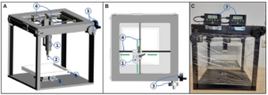
This device is the result of the collaboration of a team of engineers and medical professionals who aim to improve the success rates of tumor removal surgeries by taking advantage of the properties of Raman spectroscopy. This device, shown in the figure below, scans the removed tissue, collects the Raman spectra, and based on the data, generates plots that describe tumor probability as a function of resection distance. This method provides an estimate of the thickness or margin of healthy tissue surrounding the tumor. Typically, margin assessment is performed after surgery and the process can take around 8 hours to complete. However, with the development of the RIOARM device, surgeons can perform margin assessment live during surgery and take necessary action.
This device contains 3 modules: a Raman module, an optical fiber probe, and a mechanical stage. The Raman module is responsible for processing and analyzing the data while the optical probe locally measures the scattered light. The mechanical stage controls of the probe and its depth as it penetrates the tissue. It is important to recognize that the successful operation of the RIOARM device is owed to optical fiber probes. These one-time-use probes guide the light source to illuminate the tissue and then collect the scattered light. The optical fiber probes are made of high wavenumber optical fibers.
Importance of Optical Fibers
Generally, optical fibers can exhibit undesired nonlinear optical effects, a property of many optical materials. The nonlinear effects are capable of generating new frequencies which give rise to background signal noise in the optical fiber. However, the careful selection of the materials used to fabricate high wavenumber optical fibers helps mitigate these effects. Furthermore, these special optical fibers designed by Art Photonics, exhibit a high signal-to-noise ratio tailored for the Raman fingerprint region. This property enhances the detected Raman signals.
Results
The results of this study showed that the resection margins presented high accuracies for most of the measurements. The specificity for adequate and inadequate results exceeded 90%. The error was estimated to be less than 1 mm which is smaller than the required minimum margin of 5 mm. Additionally, the measurement time is reasonable given that 100 measurements were performed on a single sample in 15 minutes. This allows the surgeons an ample amount of time to remove more tissue if needed. The recently published paper contains additional information about the results of this study.
The final design and the prototype of the RIOARM device. The plastic foil wrapping the stage prevents tissue dehydration during the measurement process. 1 Optical fiber needle probe. 2 needle. 3 actuators. 4 guiding rods. 5 actuator to shift the specimen vertically up and down. 6 plate. 7 two monitors that read out the X and Y positions of the optical fiber needle probe.
Conclusion
Raman spectroscopy, born from Sir C.V. Raman’s discovery, has emerged as a powerful analytical tool reshaping scientific landscapes. Its evolution from early experiments to modern applications mirrors its versatility and adaptability across various fields. The unique properties of non-destructiveness, minimal sample preparation time and compatibility with different operating environments have fueled its widespread adoption, particularly in medical diagnostics and materials science. The recent strides in intraoperative Raman spectroscopy, exemplified by the innovative RIOARM device, mark a paradigm shift in surgical precision, offering real-time guidance during tumor removal surgeries. This fusion of scientific ingenuity and technological advancement underscores Raman spectroscopy’s pivotal role in reshaping medical interventions and scientific discoveries. As it continues to evolve, Raman spectroscopy stands poised to revolutionize further, unlocking new frontiers in diagnostics, treatments, and scientific understanding.
This article is made possible by the sponsorship of art photonics GmbH, a leading designer and manufacturer of optical probes and fiber optics solutions


