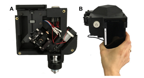
The smartphone confocal microscope could be the key to diagnosing diseases in areas with limited resources.
Researchers from around the world came together to develop a low-cost smartphone confocal microscope for imaging human skin in vivo. The team’s main concern: disease diagnosis in low- and middle-income countries.
Accurate diagnosis of diseases such as melanoma or basal cell carcinoma demands an in-depth view of the skin. Current methods of cellular-level examination (such as biopsy) require resources unavailable to lower-income countries. Additionally, lower-cost imaging methods like dermoscopy only examine the surface of the skin. This can often lead to misdiagnosis.

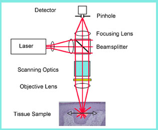
The above schematic shows how confocal microscopy can image skin tissue. Courtesy of Medgadget.
The approach
The concept of the research team’s design relies on reflectance confocal microscopy (RCM). This non-invasive technique creates a high-resolution image by viewing backscattered light from illuminated tissue. RCM has proven to provide quick and accurate results, but the complex optical setup can be expensive.
In the interest of cost, the researchers incorporated spectrally encoded confocal microscopy (SECM) into their prototype. SECM is a RCM technology which illuminates a fine line of light over the skin using a diffraction grating and broadband light source. Researchers can also use a slit aperture to form two-dimensional images without any beam scanning devices. The slit approach uses an inexpensive LED and an imaging sensor to acquire images.
This is where the brilliance and novelty of their device comes into play. Smartphone cameras use CMOS imaging sensors with a significant number of pixels and high sensitivity. Their portability and accuracy render them ideal in a SECM system. With that, the team devised a smartphone confocal microscope.

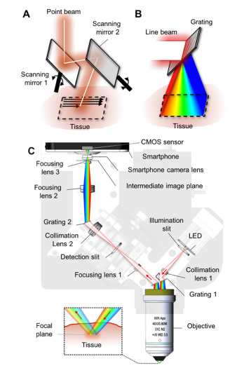
The above graphic shows the optical path within the smartphone confocal microscope. Courtesy of OSA Publishing.
Smartphone confocal microscope design
Light from a large bandwidth LED filters through an illumination slit and collimates before entering a diffraction grating. The separated beams then focus onto the skin. Reflected light reenters the objective and grating to form a single line beam. This beam focuses onto a detection slit where confocal optical sectioning takes place.
Light filtered by the detection slit diffracts one more time before entering a series of lenses to create a two-dimensional confocal image. Finally, the CMOS sensor translates the image for viewing on a given smartphone.
Image acquisition through the confocal microscope
The team took further advantage of the smartphone by using an Android application called Open Camera. This way they were able to control exposure time, image resolution, and ISO. The application also allowed them to manually control the focus of the smartphone camera to attain the sharpest image contrast.
Images were acquired in vivo and taken at multiple depth levels with motorized axial scanning.

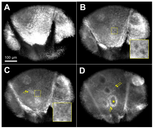
The acquired images show the skin at different depths and the yellow arrows point out basal cells. Courtesy of OSA Publishing
The results
The team successfully gathered cellular images within the human forearm. Different cell types can be viewed at different depth levels within the skin. Basal cells, which are especially susceptible to cancer, can be clearly examined at 50 micrometers under the skin surface. The researchers were also able to record video of the manual scanning process. The video shows that it is possible to image and examine different depth levels in real time.
According to the team’s paper, the resolution of their smartphone confocal microscope is comparable to that of commercially available confocal devices.
The future of the confocal microscope
Portable, cheap and powerful, the smartphone confocal microscope has the potential to revolutionize disease diagnosis in low-income countries. The total material cost is $4,200.
The team admits there are still barriers to be addressed in adapting the device in low-resource areas. Many of these challenges can be solved with additional preparation. Intermittent power supply, for example, could make it difficult to charge the smartphone. For now the 12V battery can power the LED for 11 hours. The team suggests bringing multiple rechargeable batteries to the study site. More importantly, the confocal images are useless without review for diagnosis. The researchers say the next step is to devise a system to wirelessly transfer the images to a trained professional. That person can then examine, make their diagnosis, and communicate with local staff in real time.
The smartphone confocal microscope presents a cheap and easy way to attain detailed two-dimensional images of cells. Early diagnosis is crucial, especially in areas with limited resources. Soon, this brand new confocal microscope could save lives.
For more information on the research team’s novel imaging device, click here.

I want to know more about this product… Is this confocal microscope available in market?