Introduction
Photonics has a wide array of applications in data communications, imaging, light sensing, and surgery. One such field where the utilization of light is particularly useful is ophthalmology. Photonics in ophthalmology can be used to diagnose and treat different types of eye disorders.
Using Photonics for Measurements in Ophthalmology
One example of photonics being used in this field is for the measurement of oxygen in the bloodstream; specifically, measuring the amount of hemoglobin in the eyes of patients. Hemoglobin is a protein in the blood that carries oxygen around the bloodstream. A hemoglobin count is an important indicator of whether or not the tissue is receiving enough oxygen. Essentially, how this device works is by shining lights of various wavelengths through the specific region being measured. Typically, hemoglobin that carries oxygen absorbs light at a wavelength of 660 nanometers, while deoxygenated hemoglobin absorbs light at a wavelength of 880 nanometers. This measurement is done by an optical sensor, which tracks how much light and what wavelengths are transmitted through a substance. This can help show which type of hemoglobin is more concentrated in the blood, and hence, how much oxygen is moving throughout the eye.
This sensing can be useful as in some cases there can be issues regarding a lack of oxygen associated with the eyes, such as Corneal Hypoxia. Corneal Hypoxia can bring a range of symptoms, starting with blurry vision, itchiness, and the feeling of sand in the eye, eventually leading to swelling and death of epithelial (outer eye) cells in more severe cases.
The use of photonics technologies can help identify lack of oxygen in the eyes, which may occur in the case of Corneal Hypoxia. Also, in a study done to help further the identification and observable physical effects of various eye disorders such as Glaucoma, age-related macular degeneration, and diabetic retinopathy, researchers turned to the power of photonics to help gather valuable data (specifically by measuring the retinal blood oxygen saturation rate). Essentially, photonics can be used to diagnose various ophthalmic disorders in a non-painful, non-invasive manner.
Optical Coherence Tomography in Ophthalmology
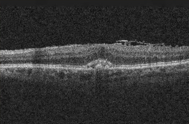
Another type of technology that can aid ophthalmologists is referred to as optical coherence tomography (OCT). Optical coherence tomography takes high resolution images of the eye, specifically the retina. Once images are taken, they can help provide ophthalmologists with images of abnormalities within the eye. OCT scanning functions by illuminating the retina with a broadband light source. The light then travels through the eye, bouncing and reflecting off the different tissue layers. The interference of back scattered light with the reference arm allows for the reconstructing the image of the eye. Below is an actual scan taken via the OCT method.

An image of an OCT scan. Courtesy of Wikimedia Commons.
SWAP Test
In addition to OCT, other photonics techniques are used in order to test for different eye problems. For instance, there is the Short-Wavelength Automated Perimetry (SWAP) test. This test is typically used to diagnose Glaucoma, which is a disease that is represented by a damaged optic nerve. This test works by putting two short-wavelength visible light sources in contrast with each other. How the patient reacts and responds to the various thresholds can show early onset of Glaucoma. The harder it is to see these lower wavelengths of light, the more likely it is that the patient has a condition like Glaucoma.
Surgical Applications of Photonics in Ophthalmology
After examining multiple ways photonics is used to diagnose disease, let’s examine other applications within the field of ophthalmology. This time, let’s review the use of different lasers that are used to perform or assist various surgeries, such as refractive eye surgery.
Typically, refractive eye surgery is used to correct various vision problems. It works by coupling different surgical techniques to restore vision. It can cure astigmatisms, nearsightedness, or farsightedness. Let’s run over a common type of refractive eye surgery, and how this surgery requires the use of photonics.
A common procedure is called LASIK. LASIK is a well known and highly effective way to battle degradation of vision. It works by utilizing lasers for surgical purposes. The procedure requires different steps. First, the doctors will calculate how much of your cornea needs to be sliced in order for your reshaped eye to have perfect vision. Once they determine that amount, the doctors will use two different types of lasers to perform the procedure.
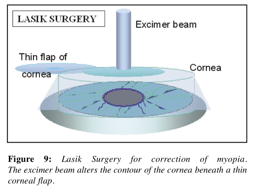
First, they use a femtosecond laser to cut a flap of corneal tissue open so that the tissue underneath is exposed. A femtosecond laser is preferable to the old method of cutting open the cornea with a blade, because the femtosecond laser “blade” cuts at an extremely fast rate, which helps avoid mechanically or thermally induced damage to the tissue. This method is much more accurate than a surgeon cutting by hand.

Courtesy of Wikimedia Commons.
Once the flap is open and the corneal tissue underneath is exposed, the surgeon will now move onto the reshaping process. Instead of using the previous femtosecond laser, they now move onto the Excimer beam, which is used to reshape the eye to allow the vision to be corrected. An excimer beam is a beam that has a wavelength of 193 nm, placing it in the ultraviolet light range. This specific type of laser is used because it has the benefit of removing parts of the cornea without much heat transfer, keeping the eye safe.
Conclusion
The field of ophthalmology and optics are deeply intertwined, leveraging the precise applications of light for groundbreaking progress. Utilizing an array of advanced technologies, such as swept lasers, which complement the rich tapestry of photonic tools, enhances the diagnosis and treatment of eye conditions beyond what was possible with traditional methods. It is anticipated that the evolution of photonics in eye care will continue to surge, with companies investing in the expansive potential of lasers and light. The future holds a spectrum of possibilities for novel therapeutic and diagnostic advancements, assuring that the role of photonics in ophthalmology will be as integral and innovative as ever.
This post was brought to you by RPMC Lasers - supplier and distributor of semiconductor lasers and laser systems.
If you are interested in reading more about related topics, click the links below:
- Laser Beam Analysis for Improving the Results of LASIK
- Optical Coherence Tomography
- Photonics and Medical Sensing
Sources:
- https://www.liocny.com/blog/corneal-hypoxia-dangers-of-oxygen-deprivation#:~:text=Symptoms%20of%20oxygen%20deprivation%20in,cornea%20and%20temporary%20blurred%20vision.
- https://www.mdpi.com/journal/photonics/special_issues/Visual_Optics_Ophthalmology
- https://parkslopeeye.com/what-is-optical-coherence-tomography-oct/#:~:text=How%20Does%20OCT%20Work%3F,3D%20image%20of%20the%20eye
- https://www.healio.com/news/ophthalmology/20120331/excimer-lasers-ushered-in-prk-and-lasik-revolutionized-refractive-surgery#:~:text=An%20excimer%20laser%20beam%20is,pain%20and%20shortens%20recovery%20time.
- https://www.ipgphotonics.com/en/applications/medical-2/ophthalmology
- https://medlineplus.gov/ency/article/007018.htm#:~:text=LASIK%20uses%20an%20excimer%20laser,the%20cornea%20to%20be%20thinner.
