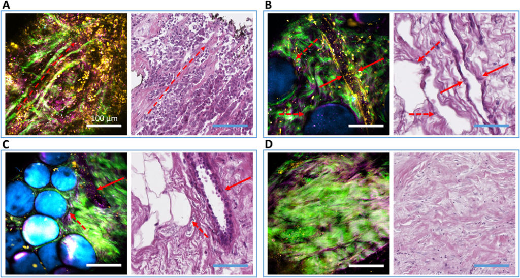
A novel nonlinear imaging system successfully captured cellular features in human breast tissue while characterizing the extracellular vesicle population. Courtesy of Science Advances.
Developing a clear characterization of a tumor’s microenvironment is imperative in understanding its progress. A new approach to determining cancer’s development involves a thorough study of extracellular vesicles (EVs) in the tissue. According to recent findings, these tiny structures that promote cell-to-cell communication in the tumor’s microenvironment could have a crucial role in the spread of cancer. Researchers hope that by efficiently quantifying the density of EVs in biopsied tissue they can significantly cut down on the labor and time typically required for diagnosis.
The Need for Faster Diagnosis
Cancer diagnoses can take several days to complete. Standard procedure requires surgically removed tissue to be processed in chemical dyes and then sent to a pathologist for extensive examination. A research team at the University of Illinois Urbana-Champaign, however, believes their device could preserve vital diagnostic information lost during this lengthy procedure. Their portable nonlinear imaging system takes advantage of nonlinear phenomena to produce high-resolution multimode images in real-time. The team successfully imaged the distribution of EVs in human breast tissue without leaving the operating room.

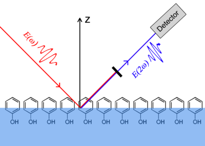
Second and third-harmonic generation can be manipulated to provide high-resolution images of biological materials. Courtesy of Wikipedia.
Nonlinear Imaging and Optics
Nonlinear optics has been one of the most versatile branches of optics since the construction of the laser in 1960. Light’s nonlinear response in certain media gives rise to a wide variety of phenomena including second-harmonic generation (SHG). SHG is commonly exploited in many medical applications due to its coherent nature. SHG microscopy is now an established method for visualizing functions and structures of cells and tissue because researchers are able to attain high-resolution, high-sensitivity images without damaging their biological sample. In addition, third-harmonic generation (THG) can provide extremely high-contrast visuals where possible. THG is exceedingly more rare than SHG as optical materials have strict phase-matching constraints for the χ(3) nonlinearity.
The Portable Multimodal Nonlinear Optical Imaging Device
The portable imaging system uses four nonlinear imaging modalities. The device uses a fifty-five femtosecond laser source with a spectral range capable of exciting the four nonlinear processes with high efficiency (1040 to 1100 nanometers). Four bandpass filters separate the nonlinear optical signals so that they are detected in different spectral windows. This form of multimode imaging allowed the researchers to target features of interest concurrently. Finally, the imaging system sits in a portable cart so that it can be easily moved within hospitals and operating rooms.
The team imaged and analyzed breast tissue from 29 breast cancer patients and 7 cancer-free participants for comparison. The portable design allowed the researchers to image the cancerous tissue in the operating room during surgery. Each analyzed site was chosen to be closest to the tumor within the specimen. Tissue sample were extracted from the healthy patients during breast-reduction surgery. As a testament to the efficiency of their device, all multimodal images of the undisturbed breast tissue were acquired within less than thirty minutes of extraction.

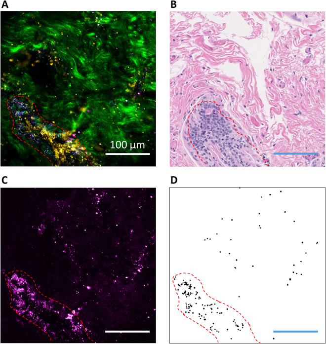
The above figures show the results of the nonlinear imaging process. Here we see a site with ductal carcinoma in situ (a) and the relevant binary image of EVs (d) on a 100 micrometer scale. Courtesy of Science Advances
THG and Extracellular Vesicles
The research team relied mostly on THG to image the EVs. They then ran these images through an automated segmentation algorithm to isolate the structures from their background and quantify EV density in both the cancerous and cancer-free samples. As expected, the research team saw a clear increase in average EV count between the two groups. Cancer cases averaged around 142/nl where healthy cases averaged 23/nl. Furthermore, EV increased with previously diagnosed grade. That is, patients with higher stage breast cancer have more aggressive tumor growth and this is reflected by the EV count.
Multimodal nonlinear imaging allows the research team to image and analyze extracted human breast tissue during surgery. The research team confirmed the accuracy of their device by comparing healthy and cancerous breast tissue. They also supported the theory of EV count by comparing different stages of breast cancer. The portable multimodal nonlinear imaging device could become common practice in operating rooms worldwide. The team believes their instrument could work with samples and procedures beyond this study. Soon this method could work with outpatient procedures or other biological samples including blood and urine.
For more information on their study and portable imaging device, refer to the research team’s paper published in Science Advances.
