In healthcare terminology, diagnostics refers to classifying an illness or condition based on studying the symptoms. The process of diagnostics has been forever changed with technology. Quality medical information from clinics, specialists and academic journals is regularly posted online for use by the public. Certain services can even be distributed through smartphone apps or other mobile technologies (Health app on iOS, or the Fitbit). However, laboratory diagnostics is a much tougher problem to address with mobile technology. This is due to the need for large, cumbersome and expensive tools for microscopy. Which is why developers want to make more nuanced services available with smartphone technology.

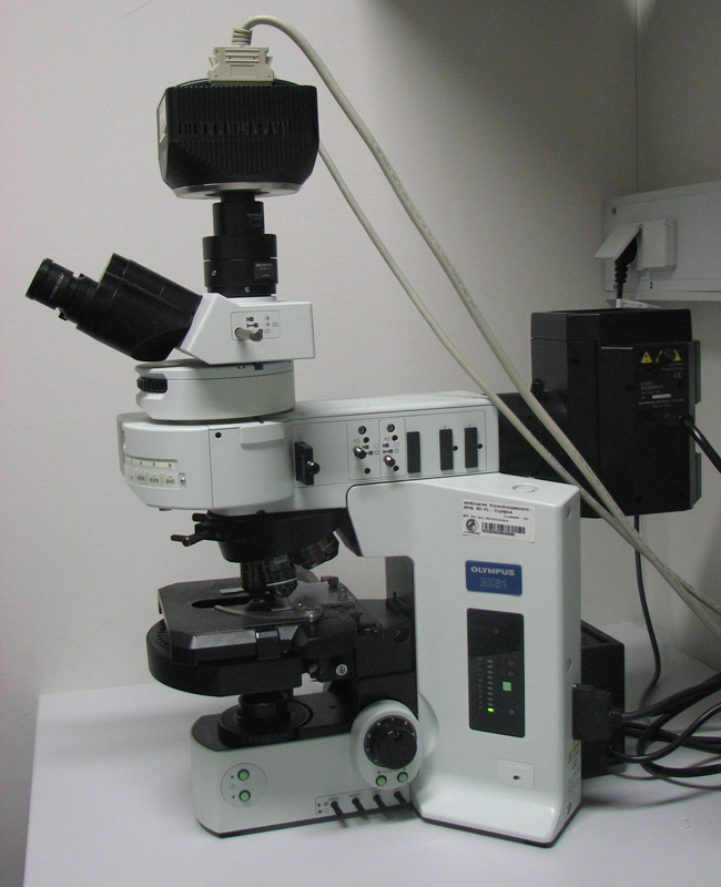
Laboratory grade microscope for fluorescence microscopy. Obviously, it is not portable and has many different parts. Overall it is difficult to adjust and requires training for operating know how.
To be clear, the focus on smartphones for this purpose is partly due to its mass adoption. Smartphones already have computation power, connectivity, and exceptional cameras in addition to being relatively easy to access. This has incentivized research into smartphone camera’s ability to replace other imaging solutions. More specifically, many different forms of microscopy like common bright field, fluorescent and even phase contrast microscopy are practically feasible with the use of commonly available smartphones.
On the other hand, microbiology studies and in-vitro diagnostics using microscopy are vitally important to studying diseases that are rampant in developing nations, but require specialized personnel to operate. While the reliance on legacy tools for these diagnostic techniques limits their use in many parts of the world, researchers are working hard to bridge this gap by simple annexations (e.g. lenses) to smartphone cameras.
Simple Bright Field Microscopy with a Smartphone
The problem that lies with many of the more complex lens devices is that they too are difficult to assemble and install on the camera. They use external light sources which results in a more bulky and expensive attachment. Also, external light sources entail the need for external batteries or increased smartphone battery usage, which leads to an additional cost. If the attachment over the lens is just as cumbersome and difficult to prepare as a lab microscope, it defeats the purpose. This is unfortunately the case for many of the early experiments with smartphone microscopy.
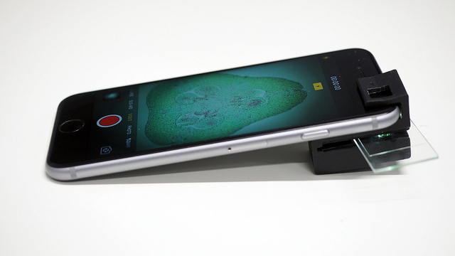
Inventors have addressed the issue of external light for bright field or dark field microscopy in smartphones. They use a 3D-printable microscope clip that fits over the camera. Inside the clip are illumination tunnels that amplify the smartphone’s flash to an intensity appropriate for microscopy. In fact, the internal reflection that occurs in these tunnels allows enough ambient light to be used in lieu of a flash. This enables the use in basic smartphones that lack the bright flash of newer ones. Additional LEDs are no longer necessary for basic bright and dark field microscopy.

Pictured above is the smartphone clip that enables light field microscopy. It is highly compact, and uses a simple, low cost lens. Picture retrieved from Flickr, image courtesy of CNBP/cnbp.org.au.
While 3D printing technology is not cheap, there is only one part that needs to be produced for this product. It is likely much cheaper to produce one clip that fits over a smartphone lens, than to invest in a laboratory grade microscope. No additional attachments are needed, and the smartphone camera brings ease of recording microscopy. iPhone’s autofocus feature, and various camera apps for iOS and Android can help with fine tuning the focus. These features are an important step in making cellular study more accessible. However, light and dark field microscopy comprises only a fraction of the market’s needs. As people experiment more and more with smartphone setups, they will find easier methods applicable to more advanced purposes.
Fluorescence Microscopy on a Smartphone
Researchers are experimenting with do it yourself methods of fluorescence microscopy with a smartphone. Fluorescence microscopy is used to identify molecular components in biology and materials science. To see sub-cellular objects requires a much more complex setup than light field microscopy. For this reason, it is much less accessible to the public. The idea is to keep the assembly simple and cost effective so that it can be proliferated.
For fluorescence microscopy on the smartphone, it is difficult to avoid using external LEDs. These measurements need much stronger light sources and can’t get by with just the flash or ambient light. Most forms of portable fluorescence microscopy require positional adjustment of the LEDs for different lenses of different strengths. Again, these forms are more difficult to assemble and operate which removes the “do-it-yourself” aspect. Much like the light field smartphone microscope that was previously discussed, this new lens harnesses total internal reflection. With this, the microscope does not need directional adjustment of the LED lights. Unlike the light field lens, this tool requires 5 parts produced with a 3D printer.
While this may sound like a lot to assemble on a smartphone, it is significantly easier and less expensive than laboratory fluorescence microscopes. The entire setup costs less than $20 to produce in a 3D printer and weighs about 26.5 grams, making it a good portable solution. It is designed for a Lumia 640, which is one of the cheaper smartphones on the market. It includes an adapter for the smartphone, multiple lids and covers to keep out ambient light and a piece to hold a glass slide. The 5-part configuration allows for customization, as parts can be replaced or altered for different purposes. For example, this same config can be used for bright field microscopy when the LEDs and lids are removed. The adapter can also be modified for use in a multitude of smartphones.
Conclusions
Smartphone microscopy techniques are still early on in their development, but they can change diagnostics for the better. The methods of smartphone microscopy provide adaptability that is most desperately needed for people without access to sophisticated laboratories. Developers have ensured the 3D print methods are open source and available online. Parts in these devices are interchangeable which allows them to be used with lenses and smartphones of varying cost and quality.
It may seem that microscopy is a luxurious tool that doesn’t have use other than abstract laboratory research. However, this couldn’t be further from the truth. Water resources in developing countries are not as reliable and need closer study to identify parasites. Crop illnesses can ruin the livelihood of generations if they aren’t found quickly, since pesticides and treatments are not so widely accessible. Allowing smartphones to perform microbiological studies could make it much easier for people in developing nations to find and study these problems. Subsequently, they can innovate, and find unique solutions to the problems they face.
Further Reading
A. Orth, E. R. Wilson, J. G. Thompson & B. C. Gibson, “A dual-mode mobile phone microscope using the onboard camera flash and ambient light,” Scientific Reports volume 8, Article number: 3298 (2018) https://doi.org/10.1038/s41598-018-21543-2
Yulung Sung, Fernando Campa, and Wei-Chuan Shih, “Open-source do-it-yourself multi-color fluorescence smartphone microscopy,” Biomed. Opt. Express 8, 5075-5086 (2017) https://doi.org/10.1364/BOE.8.005075
